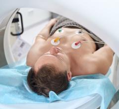
Photo courtesy of Toshiba
The application of computed tomography in cardiovascular practice has utilized electron beam computed tomography (EBCT) and multidetector computed tomography (MDCT). Both are useful for calcium scoring; however, the superior spatial resolution of MDCT has made it the dominant modality in arterial imaging.
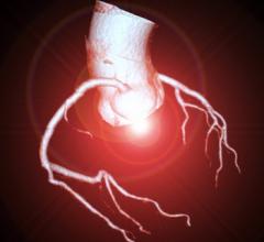
Current generation MDCT has submillimeter spatial resolution (0.75 mm) and improved temporal resolution (50 to 200 ms) as compared to earlier models. It can accurately define coronary arterial and venous anatomy, and the dataset can be acquired with a one- to 10-second breath-hold. Since the image quality is dependent on heart rate, patients are pre-medicated to attain a heart rate less than 65. After processing these images in a dedicated workstation, axial images can be reformatted into curved multiplanar reconstructions, maximum intensity projection and volumetric 3-D reconstruction.
Coronary MDCT can visualize both calcified as well as noncalcified plaque. Sensitivity for detecting coronary artery stenosis varies from 86 to 95 percent and specificity 95 to 98 percent.1 As the calcium score exceeds more than 500, the specificity of the study decreases and should not be performed. Most studies report high negative predictive value in the range of 96 to 99 percent.
Since bypass grafts are larger in diameter and have less intrinsic motion, they are readily visualized. MDCT is highly sensitive for delineating the entire length of thoracic aorta for the diagnosis of aortic dissection as well as aortic aneurysm. It is frequently used for “triple rule-out,” for example, coronary artery disease, aortic dissection and pulmonary embolism. In addition to coronary arteries, cardiac CT can also delineate disorders of pericardium (pericardial calcification, pericardial thickening, pericardial effusion), cardiac masses, valvular structure, wall motion and ejection fraction.
The current paradigm of cardiovascular disease management is that the intensity of intervention should be adjusted to the level of the baseline risk. It has been well documented that a significant majority of myocardial infarctions and acute coronary syndromes (ACS) occur at the site of previous mild or moderate stenoses due to plaque rupture. As conventional coronary angiography can only assess the lumen, it is a poor predictor of future events. In addition to visualizing the lumen, MDCT can be used to assess the vessel wall and to distinguish between calcified plaque and noncalcified “soft” plaque. Software packages for plaque analysis are currently being validated.
The value of CAC scoring
The extent of coronary atherosclerosis is an important predictor of ACS. Coronary artery calcium (CAC) is virtually always associated with atheromatous plaque.2 Though calcified plaque is stable in that it does not usually result in unstable syndromes, its co-occurrence with noncalcified plaque provides the means for estimation of cardiac risk.3
CAC has been correlated both in histological4 as well as IVUS5 studies of combined calcified and noncalcified plaque. CAC provides an estimate of total coronary plaque burden, which is independently predictive of coronary heart disease (CHD) outcome above and beyond traditional historical and measured risk factors, C-reactive protein, family history of coronary artery disease (CAD) or BMI. A score of below 10 is considered minimal and more than 400 is considered severe.
Asymptomatic patients in the 10 to 20 percent Framingham 10-year risk category (NCEP III — moderately high risk) are good candidates for CAC scoring.6 In this population, CAC testing provides independent, incremental prognostic value. If these patients have a CAC score of more than 400, then they would be expected to have CHD event rates greater than or equal to 20 percent (CHD risk equivalent). These patients can become candidates for aggressive LDL reduction toward secondary prevention goals. In certain instances, scanning low risk patients (FRS
CAC scanning may also have utility in patients with a Framingham high-risk category. For instance, some Framingham high-risk patients may be intolerant of statins or may strongly prefer alternative medicine. In these patients, CAC evidence of high risk may be utilized to reinforce the necessity for finding a statin that can be tolerated. Conversely the absence of significant CAC may permit relaxation of the treatment goals.8
Though repeated scanning is not currently recommended, Raggi et al has shown that CAC progression is greater in patients with future MI, and progression of the score was associated with a worse prognosis when compared to stabilization, regardless of the baseline level. There is currently no available data to show that aggressive lipid control will slow progression of CAC.
Progression of CAC was significantly greater in patients receiving statins who had an MI compared with event-free subjects despite similar LDL control. A CAC of 0 is associated with a low annual event rate of 0.11 to 0.12 percent.
The main disadvantages of MDCT are the need for ionizing radiation, use of contrast media, poor resolution of distal vessels, inadequate imaging in the presence of significant calcification and motion artifacts. Despite these limitations, widespread availability, plus the noninvasive nature and short exam time will enable MDCT to be the modality of choice for both diagnosis as well as follow-up of CAD. With the development of multidetector scanners and nonionic contrast material, this technique has a safety profile that clearly exceeds that of conventional angiography.
Dr. Craig Walker is founder and medical director of Cardiovascular Institute of the South, Houma, LA. Dr. Walker is author or co-author of more than five dozen medical publications and has delivered scores of lectures to both lay and medical audiences. He maintains the distinction of being a primary investigator for several cardiovascular and peripheral devices. He is also a member of the editorial advisory board of Diagnostic & Invasive Cardiology magazine.
References
1. Raff GJ, Gallagher MJ, 'Neill WW, Goldstein JA. Diagnostic accuracy of noninvasive coronary angiography using 64 slice computed tomography. J Am Coll Cardiol 2005;46:552-7
2. Rifkin RD, Parisi AF, Folland E. Coronary calcification in the diagnosis of coronary artery disease. Am J Cardiol !979;44:141 -7
3. Greenland P, Bonow RO, Brundage BH, et al. ACCF/AHA 2007 Clinical Expert Consensus Document on Coronary Artery Calcium Scoring by Computed Tomography in Global Cardiovascular Risk Assessment and in Evaluation of Patients With Chest Pain. A Report of the American College of Cardiology Foundation Clinical Expert Consensus Task Force (ACCF/AHA Writing Committee to Update the 2000 Expert Consensus Document on Electron Beam Computed Tomography). Developed in Collaboration With the Society of Atherosclerosis Imaging and Prevention and the Society of Cardiovascular Computed Tomography. Circulation 2007
4. Rumberger JA, Simons DB, Fitzpatrick LA, et al. Coronary artery calcium areas by electron beam computed tomography and coronary atherosclerotic plaque area: a histopathologic correlative study. Circulation1995;92:2157- 62
5. Baumgart D, Schmermund A, Goerge G, et al. Comparison of electron beam computed tomography with intracoronary ultrasound and coronary angiography for detection of coronary atherosclerosis. J Am Coll Cardiol 1997;30:57 - 64
6. Hecht HS, Budoff MJ, Berman DS, Ehrlich J, Rumberger JA. Coronary artery calcium scanning: Clinical paradigms for cardiac risk assessment and treatment. Am Heart J 2006;151:1139-46.
7. Pohle K RD, Ma¨ffert R, et al. Coronary calcifications in young patients with first, unheralded myocardial infarction: a risk factor matched analysis by electron beam tomography. Heart J. 2003;89:625-8.
8. Harvey S. Hecht, M., FACC,a Matthew J. Budoff, MD, FACC,b Daniel S. Berman, MD, FACC,c,d,e James Ehrlich, MD,f, M. and John A. Rumberger, PhD, FACCg New York, NY; Torrance and Los Angeles, CA;, et al. (2006). "Coronary artery calcium screening: Clinical paradigms for cardiac risk assessment and treatment." Am Heart J 151: 1139-46
9. Raggi P, Callister TQ, Shaw LJ. Progression of coronary artery calcium and risk of first myocardial infarction in patients receiving cholesterol-lowering therapy. Arterioscler Thromb Vasc Biol 2004;24:1272-7

 February 06, 2026


