
An example of the newest generation of smart cardiac CT software that automatically identifies the anatomy, auto-traces the centerlines on the entire coronary tree and labels each vessel segment. This greatly speeds CT workflows, saving time for techs, radiologists and cardiologists.
Here is a round-up of the latest technology updates and trends in cardiac computed tomography (CT) from the Society of Cardiovascular CT (SCCT) 2018 meeting in July.
Trends and Advances in Cardiac CT Technology
I spoke to several thought leaders about what is new and exciting in cardiovascular CT technologies. I caught up with outgoing SCCT President Todd Villines, M.D., FACC, FAHA, FSCCT, director of Cardiovascular Research and Cardiac CT programs at the Walter Reed National Military Medical Center, and he outlined the main tech trends in a VIDEO interview. These included:
• Artificial intelligence (AI) integration across vendors to aid quantification and workflow and advanced algorithms to pull more diagnostic data out of CT image datasets. Villines said deep learning will maximize efficiency. This includes automatic iso-centering of patients on the scanner, automated identification of anatomy, specific coronary vessel segments, plaque and calcium detection.
• Use of CT to guide interventions. Villines said this includes use of CT imaging for structural heart procedure patient selection, planning access routes, device sizing and to guide the actual interventions. CT has become a gold standard for transcatheter aortic valve replacement (TAVR) and left atrial appendage (LAA) occlusion planning and guidance, but it plays an even more important role with the more complex transcatheter mitral valve replacement (TMVR) procedures coming into use. CT is also being used more to guide interventional procedures.
One of the top experts in the use of CT in structural heart at SCCT was Jonathon Leipsic, M.D., FSCCT, professor of radiology and cardiology at the University of British Columbia, Vancouver, Canada. We spoke about his experiences with TAVR and TMVR trials and device planning in the VIDEO: The Essentials of CT Transcatheter Valve Imaging.
Interact with a 360 photo view of Canon's TAVR CT Assessment Software
• Fractional flow reserve CT (FFR-CT), which can non-invasively derive FFR blood flow readings for the entire coronary tree using computational fluid dynamics supercomputing algorithms. This can show if intermediate or significant coronary plaques are actually flow limiting or not to definitively screen a patient to be sent to the cath lab for an intervention, eliminating the need for diagnostic catheterizations.
• CT perfusion, which maps the iodine contrast levels in the myocardium through the cardiac cycle, is starting to gain market traction thanks to validation studies showing its diagnostic accuracy compared with the gold standards of magnetic resonance imaging (MRI) and nuclear perfusion exams, Villines explained.
One of the top experts in CT perfusion is Gianluca Pontone, M.D., Ph.D., FSCCT, director of cardiovascular MRI, Centro Cardiologico Manzino, Milan, Italy. He shared his views on CT perfusion and what is needed in a scanner in the VIDEO: The State of CT Myocardial Perfusion Imaging.
• Spectral CT is likely going to become a standard part of CT imaging in the years to come, Villines said. He said the ability to look at images at different kV energy levels allows cardiologists and radiologists to see different things than on average kV range images. This includes the ability to better image myocardial perfusion deficits, aid in metal artifact reduction to see restenosis inside stents, reduce metal blooming due to pacemaker leads or surgical clips, enhance plaque imaging, direct infarct imaging and to better identify thrombus.
I spoke about the use of spectral CT with incoming SCCT President Suhny Abbara, M.D., FSCCT, chief of cardiothoracic imaging and chair of the CT operations committee, University of Texas Southwestern Medical Center, Dallas. He is also an editor of the new RSNA journal Radiology: Cardiothoracic Imaging. He spoke about his experiences with spectral at his hospital in the VIDEO: Applications of Spectral CT.
• More sensitive CT detector technology that can reduce dose and increase image resolution. Two CT vendors now offer systems with resolutions down to 0.25 mm, when the standard today is 0.5. Villines said the newest generation detectors in development use computer power and photon counting to improve image quality beyond today's generation of detectors.
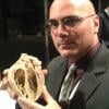
Another area of interest at SCCT were 3-D printing of cardiac CT scans for procedural planning and device sizing. Interact with a 360 degree photo showing several examples of 3-D printed heart models.
The only CT scanner on the show floor was the GE Healthcare Cardiographe, the first dedicated cardiac CT system. Interact with a 360 degree image of the CT system.
FFR-CT Gaining Momentum and Reimbursement
The use of non-invasive fractional flow reserve CT (FFR-CT) was one of the hottest topics at SCCT. The headline speaker was interventional cardiology pioneer Patrick Serruys, M.D., Ph.D., Imperial College London, who was the principal investigator of the late-breaking SYNTAX III Trial presented at EuroPCR in May. He presented the trial data again at SCCT. His comments at both SCCT and EuroPCR outlined his enthusiasm for FFR-CT, which he described as a paradigm shift in how patients with coronary disease will be assessed in the future.
In his opinion, Serruys said the technology could make traditional invasive diagnostic cine-angiography obsolete in the coming years. He said the detailed CT anatomical images showing coronary plaques, combined with the physiological information of FFR-CT showing the hemodynamic impact, can make CT a one-stop-shop cardiac imaging modality. He said it not only helps determine within a high level of certainty if a patient needs to go into the cath lab for a percutenous coronary intervention (PCI), but offers planning images so the interventionist knows exactly what they are getting into.
Additionally, Serruys said the potential for FFR-CT is much greater if it could be used as a new type of non-invasive screening test using a new hybrid Syntax score for more accurate risk assessment and early detection of disease years before clinical symptoms appear. Used in this manner, FFR-CT could become a cornerstone in a prevention strategy, where early disease detection could prompt early medical therapy intervention; follow-up imaging could confirm for the physician if the patient is compliant and visually show the patient the effect drugs have on reversing their disease.
Read more about FFR-CT presentations at SCCT in the article "FFR-CT is Ready for Prime-Time Evaluation of Coronary Disease."
Watch my interview with Serruys in the VIDEO: Will CT-FFR Replace Diagnostic Angiograms?
Kavitha Chinnaiyan, M.D., FACC, FSCCT, associate professor, Oakland University, William Beaumont School of Medicine, Royal Oak, Mich., also presented on FFR-CT. Watch her interview about her experience with the technology in the VIDEO: Using FFR-CT in Everyday Practice. Beaumont has been using the FFR-CT technology for more than three years, so it has some of the most extensive experience in the U.S.
SCCT Looks to Expand Cardiac CT Education
The society has gone through a transformation over the past year and has a renewed focus on providing educational opportunities for its members, which includes cardiologists, radiologists, radiology techs and nurses. I spoke with SCCT Education Committee Chair Ed Nicol, M.D., FSCCT, MBA, head of cardiac CT, Royal Brompton Hospital, London, who outlined new initiatives in the VIDEO: Cardiovascular CT Educational Initiatives by SCCT. This includes expanding online access to CME credit educational sessions and webinars.
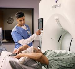
Educational tracks at SCCT include training for techs and nurses, who are on the frontline of CT scanning and interaction with the patients. I spent some time in discussion with Patricia Dickson, LRT(CT), director of imaging and outpatient services, Capital Cardiology Associates, Albany, N.Y., and Nikki Weber, a lead CT technologist at the Mayo Clinic, Rochester, Minn., to get their thoughts on how to improve CT scan quality in the VIDEO: Tips and Tricks to Aid Cardiac CT Technologist Workflow.

 February 06, 2026