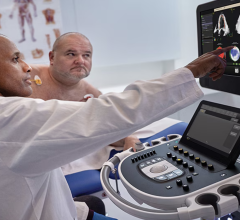
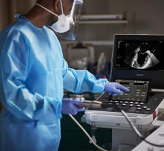
A view of GE Healthcare's volume max, or Vmax, which allows for nearly triple the frame rate speeds on 3-D TEE in a single beat over previous-generation systems.
The higher expense and lower frame rates of 3-D cardiac ultrasound systems have limited their adoption over the past decade and have begged the question at the annual American Society of Echocardiography (ASE) meetings whether 3-D is a necessary technology. However, sessions at ASE 2018 showed the focus on 3-D imaging systems has definitely changed from a research tool to a front-line echo workhorse. Clinical evidence has shown 3-D echo allows for more precise measurements, better reproducibility between operators of varying skill levels, and the technology allows for automated quantification and an increasing role for artificial intelligence to greatly speed workflows.
Most 3-D systems are still operating below 30 frames per second, but the technology and speed is improving each year, said Lissa Sugeng, M.D., associate professor of medicine, director of echocardiography and director of the Yale Echo Core Lab, Yale School of Medicine. She believes the time has come where all echo labs need at least one 3-D echo system. She said these systems are needed minimally for cardio-oncology patient assessments. The imaging they provide is also valuable for surgeons and structural heart interventionalists who need the 3-D imaging for a more comprehensive assessment and visualization of valves, septal defects and the left atrial appendage.
As the frame rates improve and advantages in workflow are better appreciated, Sugeng said 3-D will likely begin to replace older 2-D systems in the coming years.
"I think three-dimensional echo has advanced very rapidly within the last five years," she said. "We have seen developments where I think we are at a point we could live in just 3-D echo space."
3-D TEE Offers Most Advanced Renderings
With the need for real-time transesophageal echo (TEE) to guide many of the transcatheter structural heart repairs now performed, vendors have put a lot of emphasis into fast TEE to aid image guidance. Procedures like MitraClip placement would be impossible to perform without high-quality TEE.
For this reason, 3-D TEE frame rates are higher, resulting in better quality imaging.
Sugeng said 3-D echo allows 2-D slices (MPRs) to be created. This can be very helpful with sonographers that are not as experienced where an optimal 2-D view can be created in postprocessing. She said the image quality of 3-D systems was previously not as good as 2-D systems. But recent generations of 3-D echo platforms now have image quality that is on par with 2-D echo, so there might not be a need to use 2-D anymore, Sugeng explained
"I do this already," she said. "In our lab we do 3-D left ventricular (LV) volumes and left atrial (LA) volumes on a daily basis. Particularly with the new workflows some of the companies have, it's really a very smooth and rapid workflow where you can extract the three-dimensional volumes without a lot of effort."
Transthoracic Frame Rates, Volume Rates are Increasing But Behind 2-D
Frame rate speed is what makes the difference between lower quality and higher quality echo imaging to capture the details of a fast-moving heart. The issue with 3-D is instead of just setting data for one slice or multiple slices, 3-D needs to send and render much larger image volumes of data. This slows down the processing speed on ultrasound systems and some of the data needs to be ignored to make the images appear in real-time on the screen.
"When you look at finer details on 3-D transthoracic imaging, I don't think the image quality is quite there yet in replacing 2-D with 3-D echo," Sugeng explained. "They are approaching it with faster 3-D volume rates and 2-D frame rates that are higher. In biplane imaging on 3-D systems we typically see frame rates lower than 30, which is insufficient."
But she can see some of the companies pushing their technology so their frames are almost equivalent to their 2-D echo, single-plane systems, so biplane with higher frame rates are becoming available. However, when you translate that into 3-D volume rates, companies are trying to push the envelope and reach 20 and sometimes 30 volumes per second. "So, we are close in terms of temporal resolution," Sugeng said.
She said some vendors have attempted to increase their volume rates by using multi-beat acquisitions, but Sugeng said she would prefer a one-beat solution to avoid image stitching artifacts.
As computing power continues to advance, she said it is inevitable that frame rates will catch up in 3-D systems. "I think all the companies are trying to strive for that," she said.
An advancement on that front came with the 2018 release of GE Healthcare's Imaging Elevated version of its cSound image reconstruction technology. The technology aids imaging quality, workflow and quantification on the Vivid E95 cardiac imaging system. It leverages GPU processing to improve the volume frame rate, which GE refers to as volume max, or Vmax. This allows for nearly triple the frame rate speeds in TEE in a single beat over previous-generation systems.
Automation Speeds Echo Workflow
Automation has become standard on all the new echo systems, including integration of artificial intelligence. The goal of this is to speed up workflow to cut the time required for an echo exam. The machines now can perform auto-quantification in seconds that would have required several minutes of staff time in the past. This is particularly true of 3-D systems, where the machine algorithms perform auto 3-D volume assessments.
"Automation has made our workflow that much better," Sugeng explained. "There are companies now that do anatomical intelligence and deep learning that makes our quantitation of LV volumes and LA volumes much easier than before and it is actually more applicable in a routine setting rather than in a research setting."
She said this goes beyond quantification to automation to speed rendering of the image to improve workflow during procedures.
"The one thing that I would say is really important for us is to have better workflow," Sugeng said. "So if I am in an interventional procedure, I don't have time to position the valve and rotate it 90 degrees in a Z-plane. I need that valve immediately in the surgeon's view, and I need it immediately from a ventricular view, so a lot of the systems now give us the option of dual view. The workflow is very key for us because we need things in seconds."
3-D Echo Aids Structural Heart Evaluations
Three-dimensional echo is a big help for interventional cardiologists and surgeons on the heart time to better evaluate anatomy like valves. Sugeng said 3-D allows for the deconstruction of the valve. "If the image quality on 3-D echo is so superb that we can go into the data set to look at 2-D MPRs and learn a little more about the two-dimensional planes with high quality, it would be a large advantage," she said.
She said the problem with 2-D echo is that the planes are not always aligned and can misrepresent the valve scallops very easily.
She said many of the newer 3-D echo systems now offer advanced rendering algorithms to reconstruct images to greatly enhance diagnosis and procedural navigation. This includes 3-D surgical views with the ability to flip the perspective around to show the opposite side of the anatomy like on the Siemens eSie Valve software, or the ability to change the position of lighting to boost contrast of the anatomy as on GE Healthcare's Vivid E95 and on the latest version of the Philips Epiq.
"This enhances our understanding of the anatomy and issues. Take MitraClip for example, it can show you were the regurgitation jet is coming from," she said.
Advice for Labs Buying New Echo Systems
Sugeng said labs should be looking to buy at least some 3-D systems looking into the future. "I know that these are more expensive, but when you think about strategically positioning oneself, you should buy into three-dimensional echo," Sugeng explained. She said this is particularly true for cardio-oncology applications.
However, she said maybe a lab does not need to buy all 3-D systems at once, but it is good to have a couple in the lab.
Watch the VIDEO: Can We Live in 3-D Echo? — an interview with Lissa Sugeng, M.D.
Related Echocardiography Advances:
Recent Advances in Echocardiography Technology
Top Technology Trends in Echocardiography at ASE 2018
VIDEO: Editor's Choice of the Most Innovative New Echo Technology at ASE 2018
ASE Releases New Guide to Performing Comprehensive Transthoracic Echo Exams
360 View of EchoPixel's true 3-D Visualization Console at ASE 2018
Cardiovascular Ultrasound Technology Showcased at ASE 2018
VIDEO: 4 Recent Advances in Echocardiography Technology — Interview with Sunil Mankad, M.D.
VIDEO: Artificial Intelligence in Cardiac Ultrasound — Partho Sengupta, M.D.
360 View of a GE E95 Echocardiography System at ASE 2018
Recent Advances in Echocardiography Technology
360 Photo of Philips Epiq Cardiac Ultrasound System at ASE 2018
VIDEO: Tricuspid Valve Imaging and Interventions Developing Hand-in-hand — Interview with Rebecca Hahn, M.D.

 June 15, 2026