Magnetic navigation technology to direct and digitally control catheter and guidewire devices along complex paths within the heart and coronary vasculature has been evolving since 1990, and magnetic navigation was first employed in a cardiac clinical trial in early 2001 at Barnes-Jewish Hospital in St. Louis.
But in 2003, magnetic navigation really began to come of age and is now being used in more than 40 cardiac cath labs worldwide for both electrophysiological and intracoronary applications.
Of the 30 installations in the U.S., 45 percent are in EP rooms, 17 percent are in PCI rooms and 38 percent are in combo rooms. The promise of reducing procedure times, radiation, contrast and device usage is now being realized. These all result in increased throughput in the cardiac laboratory and decreased costs on the hospital’s bottom line.
Magnetic navigation for directing and controlling devices in cardiovascular applications was developed by Stereotaxis Inc. (www.stereotaxis.com), which was formed in 1990 to further develop the pioneering work of several researchers at the University of Virginia, the University of Iowa and the University of Washington in magnetic instrument guidance. The current system is the Niobe Magnetic Navigation System.
Here’s how it works:
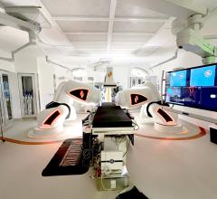
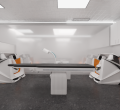
Two magnets, external to the body, control magnetic-tipped catheters and guidewires. Two computer-controlled permanent magnets, one on either side of the patient, are used to orient the tip of these specially designed devices. The magnets allow 360-degree rotation of the tip. The current Niobe software provides a touchscreen interface to guide the catheter remotely within the heart and the coronary arteries. The two magnets are tracked and swing into a fixed position with the touch of a button, and they are stored out of the way when not in use.
Human clinical trials employing magnetic navigation were first initiated in 1998 for neurosurgery applications. Cardiac electrophysiology clinical trials were done in 2001, and in 2002 the first generation navigation system, the Telstar, was cleared for market in the U.S. with a Stereotaxis-developed biplane flat panel digital imaging system.
Ablation Application
The second generation magnetic guidance system, the Niobe System, was cleared to market in the U.S. in January 2003. Niobe was first integrated with Siemens Medical Solutions' digital fluoroscopic imaging system, the AXIOM Artis dFC (flat panel system), in early 2003. The Niobe System has since been integrated with the Philips Allura XP FD (flat panel system) family as well. Stereotaxis has also partnered with Biosense Webster Inc., a Johnson & Johnson company, to integrate the Carto XP and EP Navigation and Ablation system with the Niobe Magnetic Navigation System.
In January of this year, The Cleveland Clinic, Baptist Memorial Hospital-Memphis and St. Elizabeth's Medical Center of Boston completed the first series of procedures treating cardiac arrhythmias using the Celsius RMT Diagnostic and Ablation Catheter. The proprietary catheter was jointly developed by Biosense Webster and Stereotaxis and was cleared by the FDA in late 2005.
Eric Johnson, M.D., of Baptist Memorial Hospital, successfully completed two AV node ablations with the Stereotaxis system in combination with the Celsius RMT Diagnostic and Ablation Catheter.
"The decreased procedure time and increased efficiency seen in our initial experiences using the Stereotaxis system in combination with the Celsius RMT Diagnostic and Ablation Catheter are rarely achieved when relying on manual ablation," said Dr. Johnson. "At a community hospital, speed and efficiency are obviously critical to the center's ability to increase patient volume, and I believe that leveraging this technology could be helpful in increasing patient flow."
Success in Troubled Cases
Trinity Mother Frances Health System (Tyler, TX), was one of the early recipients of the Niobe Magnetic Navigation System with the Siemens Artis dFC, installed in September 2003.
Roderick B. Meese, M.D., medical director, Trinity Mother Frances Heart Institute, reported on the institution’s experience both at TCT 2004 and again at TCT 2005.
At a Stereotaxis-sponsored breakfast meeting during last October’s meeting, Dr. Meese stated that theirs is a clinical arena, not an academic one. The hospital's experience with PCI is that 25 percent of cath lab patients are contraindicated for PCI1, that is, unable to be treated or unlikely to be effective. These are the patients he sees as candidates for magnetic guidance technology. Exams are performed more quickly with less radiation and contrast, and trauma to vessels is limited since softer wires are used to transverse tortuous vessels.
Examples shown at this presentation included guiding a wire around a 150-degree turn to reach a stenosis following a saphenous vein graft. Patients referred to Trinity often include those who have had an intervention fail using standard techniques. In another example, a PCI was successfully accomplished in 30 minutes with magnetic navigation on a patient where a previous unsuccessful attempt without magnetic navigation was abandoned after two hours.
Radiation Reduction
In the EP lab, the need to improve throughput and reduce radiation exposure is a key driver in the purchase decision of a magnetic navigation system. EP studies have been growing exponentially as new and extremely effective therapies have been developed over the last five years. The primary limits to growth at many labs are the number of rooms and qualified staff, making the issue of improved productivity paramount.
Gery Tomassoni, M.D., medical director of the EP Lab at Central Baptist Hospital (Lexington, KY), performed the first cardiac procedure using the Niobe Magnetic Navigation system on March 18, 2003. Central Baptist-Lexington has a total of eight cath labs, three of which are dedicated EP labs; five are general cath labs. One of the EP labs and one of the general cath labs have a magnetic guidance system. By mid-year both of the magnetic guidance systems will be dedicated to EP labs. All three of the EP cardiologists practicing at Central Baptist-Lexington wish to do the majority of their procedures employing the magnets. By comparison, the magnets are used in a minority of cases for PCI.
Since the first magnet was installed, Dr. Tomassoni and his colleagues have done over 550 EP studies employing magnetic navigation. Most of the exams assisted with magnetic guidance have been diagnostic EP studies, biventricular lead implantation or supraventricular ablations (ATTRAC I & II trials).
However, now that the Celsius RMT Diagnostic and Ablation Catheter is available, Dr. Tomassoni sees a dramatic increase in the number of exams performed with magnetic guidance.
Prior to this, the ablations using magnetic guidance could only be performed under protocols such as the TRAC I and TRAC II or CRAFT. Now, with the new catheter and two EP rooms equipped with magnets, he sees 80 to 90 percent of ablations being done with magnets, and they perform 500 ablations each year in his labs. Only those patients needing another catheter or who are contraindicated for an exam in a magnetic field will be done conventionally.
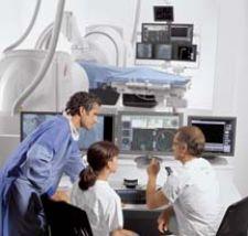
Dr. Tomassoni says the biggest advantage to the EP physician is that “the physician is out of the lab, not wearing lead or exposed to radiation.” Plus, by being in the control room, the physician is in charge of a universal master control of all of the modalities, electrical signals, Biosense, ultrasound and other functions.
The patient benefits from the reduction in fluoroscopic exposure. Since the magnet, instead of the physician’s hand, holds the catheter in place, there is “less tendency to step on the foot pedal to assure that the catheter hasn't moved slightly,” said Dr. Tomassonni.
He also believes that time will show that ablations performed under magnetic guidance will produce more consistent results and less variability between physicians. Success rates will be more consistent and complications will go down.
“The system is easy and friendly to use,” he said.
The Niobe Magnetic Navigation System can be installed in a standard cath lab measuring approximately 22 x 21 feet as shown in the installation diagram. Each magnet weighs 4,000 pounds and generates a five-gauss field.
At 1/20th the field strength of the average MRI system, the contraindicators for MRI studies do not apply for a magnetic navigation study.
However, as Jane Van Dyne discovered, the magnets do add to the construction cost and time. The director of the Cardiac Catheterization Laboratory at St. Vincent Hospital in Indianapolis says that in her lab, the floor did not have to be strengthened to handle the additional 8,000 pounds of weight, but magnetic shielding did have to be added to keep the magnetic field from interfering with equipment in adjacent rooms. The first patient studies in the hospital’s Niobe-equipped EP lab were expected to have been done late in January. The St. Vincent group anticipates dramatic improvements in exam times for its biventrular and atrial fibrillation procedures, says Van Dyne.
References
1. TCT 2004 Poster 418 Magnetic Assistance in Percutaneous Coronary Intervention: Single-Center Experience; R.B. Meese, Trinity Mother Francis Hospital; D. Clark, Trinity Mother Francis Hospital; K. Elliot, Stereotaxis, Inc.
For further information, see:
1. Gallagher PL, Angel L, Martin LQ, Tomassoni G. Magnetically-assisted remote diagnostic electrophysiology studies using a single catheter and no fluoroscopy. PACE May 2005; 31:S114.
2. Gallagher PL, Angel L, Martin LQ, Tomassoni G. Magnetically-assisted biventricular device implantation: comparison using a standard guidesheath or a “bare” wire approach without a guidesheath. PACE May 2005; 4:S4.
3. McLoney AM, Martin LQ, Angel L, Gallagher PL, Tomassoni G. The use of a magnetic navigational system in the EP lab. PACE May 2004; 284: S91.


 April 20, 2026