Wide beam reconstruction (WBR) reduces both dose and image acquisition time by 50 percent for myocardial perfusion SPECT (MPI), according to a new study published in the March-April 2011 issue of the Journal of Nuclear Cardiology. WBR is a software program by UltraSPECT for reconstructing nuclear medicine images. Researchers found that with either half the dose of Tc-99m sestamibi or half the acquisition time, WBR resulted in image quality superior to processing with today’s widely used OSEM (Ordered Subset Expectation Maximization) software. The study was conducted at St. Luke’s-Roosevelt Hospital and Columbia University College of Physicians and Surgeons in New York City.
“The escalating radiation levels of today’s advanced imaging exams is causing growing concern among the medical community and the public at large,” said Gordon DePuey, lead researcher and M.D., director of nuclear medicine at St. Luke’s-Roosevelt Hospital and Professor of Radiology at Columbia University. “There is significant pressure to minimize radiation dose, particular for MPI nuclear SPECT exams.”
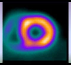
In addition, the ongoing shortages of radiopharmaceuticals due to nuclear reactor downtime compound the need to minimize dosage. WBR’s reconstruction algorithm incorporating depth-dependent resolution recovery and image noise modeling to deliver a higher quality image with lower count density data. In the new MPI study, researchers evaluated images from 156 patients undergoing myocardial perfusion SPECT with a standard full-time acquisition protocol processed with routine OSEM methods.
The same patients underwent half-time acquisition, and the data were processed with the WBR algorithm. The images were acquired both at rest and following exercise or pharmacologic stress. A second study group of 160 patients received half of the standard radiopharmaceutical dose, with images acquired for the full standard acquisition time. These were processed using WBR only. All images were rated for quality by two observers unaware of the acquisition and processing methods. For both the lower dose and abbreviated acquisition time images, grading parameters included myocardial count density and uniformity, endocardial and epicardial edge definition, visualization and definition of the right ventricle and background noise. For the abbreviated acquisition time images only, SPECT perfusion defects also were examined. Overall, WBR half-time and half-dose image quality was judged as superior to OSEM image quality in both arms of the study. There was no statistically significant difference between the two SPECT protocols in identifying the extent or severity of perfusion defects.
“The results of this study demonstrate that WBR is a powerful means of reducing dose without sacrificing image quality and diagnostic accuracy,” DePuey said. “I would recommend that all nuclear medicine laboratories adopt some strategy for reducing patient radiation exposure incorporating WBR or others techniques that have proven effective.”
DePuey notes that new SPECT cameras with multiple focused detectors are another effective dose-reduction solution. “Wide beam reconstruction technology, however, is a software-only solution,” he says. “For sites unable to budget for a major hardware acquisition, WBR provides an equally effective, more affordable answer.”
For more information: www.ultraspect.com
 June 05, 2023