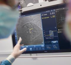
October 31, 2013 — Philips and RealView Imaging Ltd. have completed a clinical study that demonstrated the feasibility of using an innovative live 3-D holographic visualization and interaction technology to guide minimally invasive structural heart disease procedures. In the pilot study that involved eight patients and was conducted in collaboration with the Schneider Children’s Medical Center in Petach Tikva, Israel, RealView’s visualization technology was used to display interactive, real-time 3-D holographic images acquired by Philips’ interventional X-ray and cardiac ultrasound systems.
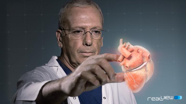
In addition to viewing the patient’s heart on a 2-D screen, doctors in the interventional team were able to view detailed dynamic 3-D holographic images of the heart “floating in free space” during a minimally invasive structural heart disease procedure without using special eyewear. The doctors were also able to manipulate the projected 3-D heart structures by touching the holographic volumes in front of them. The study demonstrated the potential of the technology to enhance the context and guidance of structural heart repairs.
“The holographic projections enabled me to intuitively understand and interrogate the 3-D spatial anatomy of the patient’s heart, as well as to navigate and appreciate the device-tissue interaction during the procedure,” said Dr. Einat Birk, pediatric cardiologist and director, Institute of Pediatric Cardiology, Schneider Children’s Medical Center.
“The ability to reach into the image and apply markings on the soft tissue anatomy in the X-ray and 3-D ultrasound images would be extremely useful for guidance of these complex procedures,” said Dr. Elchanan Bruckheimer, pediatric cardiologist and director, cardiac catheterization laboratories, Schneider Children’s Medical Center.
Bruckheimer presented the results of the pilot study at the 25th annual Transcatheter Cardiovascular Therapeutics scientific symposium (TCT 2013).
“Our ultimate goal is to create the future of healthcare by delivering innovative solutions that enhance clinical capabilities and improve patient outcomes,” said Bert van Meurs, general manager, Integrated Clinical Solutions and Marketing Imaging Systems, Philips Healthcare. “By teaming up with partners that share our passion for innovation, we have been able to demonstrate the feasibility and potential value of the world’s first holographic visualization technology targeted at guiding minimally invasive cardiac procedures.”
“I see clear indications that 3-D medical holography will play an important role in medical imaging in the near future,” said Aviad Kaufman, CEO, RealView. “With the advancement of live 3-D imaging and increasing clinical evidence of its value for a variety of procedures, we are convinced that our holographic technology will further enhance 3-D imaging and, most importantly, improve patient care.”
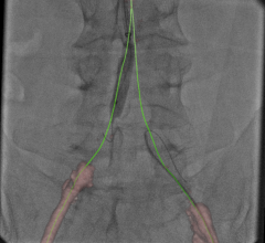
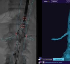
Progress in image-guided therapies for heart diseases — from the opening of obstructed coronary arteries to catheter ablation therapy for heart arrhythmias and catheter-based structural heart repairs (for example, heart valve replacements) — have greatly increased the need for live 3-D image guidance to supplement today’s live 2-D image guidance. Live X-ray and live 3-D cardiac ultrasound imaging are typically used simultaneously to guide minimally invasive structural heart repair procedures, with the ultrasound images providing detailed insights into the heart’s soft tissue anatomy and the X-ray imaging providing visualization of catheters and heart implants.
The technological advancements in the acquisition of live 3-D images to guide minimally invasive procedures have also triggered the development of novel ways to visualize the data. Following the promising results produced by this pilot study, Philips and RealView Imaging will continue to explore the clinical value of combining live 3-D imaging and medical holography in interventional cardiology and other clinical areas.
For more information: www.philips.com, www.realviewimaging.com
 January 18, 2022