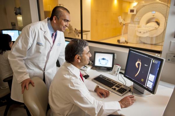
Protocols Developed for Diagnosing Pericardial Disease Using Multi-modality Imaging
August 30, 2013 — To counter the significant levels of morbidity and mortality associated with pericardial disease (disease of the sac around the heart), experts from the American Society of Echocardiography (ASE), the Society for Cardiovascular Magnetic Resonance (SCMR), and the Society of Cardiovascular Computed Tomography (SCCT) came together to review evidence and provide future guidance to clinicians. For the first time, an expert consensus statement on the appropriate use of multimodality imaging in the diagnosis and management of pericardial diseases will be published in the September issue of the Journal of the American Society of Echocardiography (JASE). The writing group was chaired by Allan L. Klein, M.D., FASE, director of Cardiovascular Imaging Research and the Pericardial Center and an echocardiographer from the esteemed Cleveland Clinic in Cleveland, Ohio.
When asked about the need for this guideline document, Klein remarked, “Pericardial disease is very common worldwide and can present in a wide range of symptoms and etiologies, making diagnosis and management clinically difficult. This document will allow clinicians in a variety of settings including primary care, emergency room and subspecialty settings, such as rheumatology, oncology and cardiology, to weigh the strengths and weaknesses of a particular imaging modality to evaluate and manage patients with suspected pericardial disease. The incremental information provided by these advanced imaging modalities may alter clinical decision making and potentially even improve outcomes.”
All patients with suspected pericardial disease, including acute, recurrent, and constrictive pericarditis as well as pericardial effusion, should have an echocardiogram as the initial diagnostic test followed by cardiac magnetic resonance imaging (MR) and/or cardiac CT, if necessary (especially in patients with poor prognostic signs such as failure to respond to anti-inflammatories), according to the guidelines. Echo emerges as the front-line imaging test used to detect most pericardial syndromes because of its ease of use, wide availability, bedside readiness, cost-effectiveness, and its comprehensive assessment of both anatomy and physiology. However, the guidelines also include a number of tables, images and other resources to guide clinicians through a stepwise approach to appropriately utilizing imaging in the diagnosis and management of various pericardial diseases. For example, CT should be used if there is calcification of the pericardium as in constrictive pericarditis.
The guidelines also emphasize the important role of inflammation in the pericardial sac often detected by cardiac MR, which then can be targeted with intense anti-inflammatory therapy. There is also an accompanying appendix, aimed at sonographers and imaging technologists, which reviews the technical approach in performing detailed “Diastology” echocardiograms, as well as the sequences and settings for cardiac MR and CT exams.
For more information: www.onlinejase.com

 June 17, 2026


