A new area of DNA testing involving telomere length may enhance a patient’s cardiovascular disease risk stratification. Telomeres are biological clocks that determine the lifespan of a cell, as well as the number of times it can divide to create replacements. Telomeres are repeating units of DNA on the ends of each chromosome that protect the information encoded in the DNA against loss. Some of these repeating units are lost with each cell division, but can be restored by enzymes in the nucleus.
During cell division, chromosomal DNA must be copied faithfully into the daughter cells. However, the ends of chromosomes are tricky to copy, and with each round of replication some of the DNA at the end of each chromosome is lost. To provide a buffer against loss of important information, the ends of chromosomes are made up of many copies of a repeating DNA sequence (TTAGGG) that is maintained independently of the normal DNA replication process, by an enzyme complex called telomerase. The gradual loss of telomeres over multiple rounds of cell division eventually lead to loss of the ability to divide further(1).
Telomere length (TL) is influenced by genetic and environmental factors. DNA variations in the genes encoding telomerase and its partners have been shown to be strong determinants of TL. Age is a powerful predictor of TL, as normal maintenance of many body systems over time requires ongoing cell replication. In addition, a variety of environmental and lifestyle stresses have been shown to accelerate telomere shortening, including emotional trauma, poor diet, inactivity and inflammation. This may be due to stress-mediated suppression of the function or expression of telomerase itself. At the same time, considerable evidence indicates that removal of harmful stresses can restore TL. A number of small studies suggest that these restorative changes may be possible over relatively short timeframes of weeks to months, and sustainable over multiple years.[2-7]
Short telomeres have been implicated in a wide range of common diseases associated with aging. Large observational studies have established an inverse association between TL and the risk of coronary heart disease (CHD) and cardiovascular disease (CVD), a composite endpoint that includes CHD, major adverse events such as myocardial infarction (MI) and stroke, and other measures of atherosclerosis progression. A meta-analysis involving 24 studies with 43,725 subjects identified a 1.54x increased risk (range 1.30-1.83) for CVD for those in the shortest third of TLs compared with the longest third, and this association appeared independent of conventional vascular risk factors.[8]
The INTERHEART study was a large, multiethnic case-control study of incident myocardial infarction, involving 3,972 subjects enrolled within 24 hours of an acute MI, and 4,321 age- and sex-matched controls without known CHD. TLs in subjects were significantly shorter than in controls (p = 3.37 × 10⁻²¹), and each unit decrease in TL increased the odds ratio of MI by 2.24 (range 1.88 to 2.67). The addition of TL to known CVD risk factors significantly enhanced the prediction model, and was independent of diabetes, physical activity, hypertension, smoking, age and ethnicity. Genetic studies further support this association.[9,10]
Mendelian randomization studies indicate that the link between short telomeres and CVD is causal. Mendelian randomization can compare populations with genetically determined differences in TL, so that the effects of TL itself on a disease process can be examined independently of varying (and presumably randomly distributed) environmental risks. Using this approach, Haycock et al. showed that longer telomeres were associated with a 22% reduction in CVD risk (11); similarly, using data from the CARDIoGRAM CHD study[12,13], Zhan found a one-unit increase in TL was associated with 21 percent reduced odds of developing CVD.[14]
How Telomere Length Influences Cardiovascular Disease
So the question is how does TL influence the development of CVD? In the study cited above, Zhan et al. concluded that insulin levels are critical mediators of this effect.[14] TL inversely correlated with fasting blood insulin levels in the multiethnic 1999–2002 National Health and Nutrition Examination Survey (NHANES), a general population survey of 7,252 adults ages 20 to 84.[15] TL was also negatively associated with adiposity, inflammation and measures of cardiac work, and positively associated with HDL cholesterol. These associations were seen across all racial and ethnic groups and in both men and women. The correlation of TL with multiple independent cardiovascular risk factors could reflect the sensitivity of telomeres to oxidative stress-related pathways downstream of each of these factors. TL thus seems to act as a readout for “allostatic load”—the summation of multiple system derangements—that can lead to heart disease.
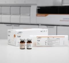
Commercial TL testing is now available from Telomere Diagnostics. The company has developed a tool, TeloYears, which is intended to help identify risk for coronary artery disease in patients 45 years of age and older who have not been previously diagnosed. Doctors can use this test to serially monitor a patient’s risk of CHD as well as their success in adhering to lifestyle modifications. The test uses quantitative polymerase chain reaction (qPCR) to measure TL in leukocytes from a finger stick blood sample, using a validated method.[16] A value is returned called the average telomere length (ATL). The ATL is calculated relative to two genes of known length that are present as single copies in the genome; this value is also referred to as a T/S ratio in published studies. The ATL is then adjusted for age and sex, and classified as long, medium or short. The classifications, based on measurements of ATL in sex- and age-adjusted peer groups from a representative sample of more than 9,300 individuals, are used to report the subject’s level of CVD risk as low, intermediate or high (with longer being at lower risk).
All TeloYears testing is conducted at a Clinical Laboratory Improvement Amendments of 1988 (CLIA)-certified laboratory at Telomere Diagnostics. CLIA certification is a rigorous process that enforces high-quality metrics, including meticulous record-keeping, quality controls, staff competency and standardized processes to assure that results are accurate and robust. The company provides extensive literature with each test kit, as well as access to medical counseling for patients with questions about their test results. Since mid-2016, TeloYears testing has been available directly to consumers via Telomere Diagnostics. TeloYears has been designed to facilitate the use of serial TL measurements by doctors who wish to provide enhanced cardiovascular disease risk assessments to their patients.
Use of Telomere Length to Aid Cardiovascular Disease Risk Stratification
Risk stratification for CHD has been intensely pursued for several reasons: CHD is highly prevalent; the time from onset to morbidity may be very brief; and in at least 25 percent of patients, CHD presents as sudden death.[17] By establishing an initial primary risk level, healthcare providers can focus their efforts and time on those patients who stand to gain the most by making significant lifestyle changes. Existing risk tools, such as the Framingham Risk Score and the 2013 ACC/AHA risk score, which sum up the impact of multiple individual risk factors, have known limitations in this regard.[18] Interestingly, the majority of people who develop CVD are not in high-risk categories: in a European study of 205,178 middle-aged individuals, more than 50 percent of CVD related deaths occurred in the subset with calculated 10-year risk of less than 10 percent.[19]
Newer parameters of risk have been extensively studied, including coronary artery calcium (CAC) scoring and determination of serum C-reactive protein; but apart from CAC, most do not greatly change the risk assessment. CAC is very useful for identifying established coronary artery atherosclerosis and for predicting incident CVD[20], but is not dynamically responsive to lifestyle changes. CAC also exposes patients to measurable amounts of ionizing radiation (although efforts to reduce this exposure are ongoing), making it less desirable for serial testing.
The TeloYears test overcomes some of these disadvantages. It can be performed serially, and reflects changes in a wide spectrum of recognized cardiac risk factors. It may also act as a biomarker for yet-unrecognized causes of CVD, such as those that drive heart attacks in patients with zero coronary calcium.[21] And the same test provides information relevant to other diseases associated with aging and allostatic load.[11]
In addition, availability of a standardized and validated test in a CLIA-approved facility can facilitate future clinical studies of TL in prospective longitudinal studies of cardiovascular risk, overcoming some of the limitations of inter-laboratory variability.[22] More clinical data accrual is needed to determine the range of subjects for which TL testing is most predictive, to show how efficiently lifestyle modification can change TL over time, and how TL determination complements or replaces more familiar cardiovascular disease markers.[23]
Future data from this test will help determine whether interventions to prevent telomere shortening affect the hard clinical endpoints of CVD. There is already substantial evidence that TL-modifying factors — such as being overweight and smoking—significantly increase CVD risk. A genetic predisposition to longer telomeres confers a reduced risk of CVD — meaning that long telomeres are themselves protective. It will be interesting to learn how much telomeres act as key defenders of cardiovascular health. Until then, the ability to slow down—or even reverse — the biological clock may provide critical encouragement for patients to adopt and commit to changes in stress levels, diet, activity level and weight.
Editor’s note: Nanette H. Bishopric, M.D., FACC, FAHA, is a professor of molecular and cellular pharmacology, medicine and pediatrics at the University of Miami (UM), a practicing cardiologist in the UM Medical Group, and director of UM Cardiovascular Genomics Laboratory. She is a regular peer reviewer for National Institutes of Health (NIH) and American Heart Association, and serves on the Editorial Boards of Circulation and Circulation Research. Her research is currently supported by NIH grants, Fondation Leducq (Paris) and Florida Heart Research Institute, focusing on molecular genetic effectors of hypertrophy, heart cell death and renewal. Bishopric's newest goal is to move the most important recent findings in this area to proof of concept in man.
Related Genetic Testing for Cardiovascular Disease Articles
Identification of Novel Cardiovascular Disease Pathways, Biomarkers
FDA Taking Genomic Testing to the Next Level
Preliminary Results Published of Largest-Ever Study Linking Biomarkers with Coronary Artery Disease
NIH Team Discovers Genetic Disorder Causing Strokes, Vascular Inflammation in Children
References:
1. Blackburn EH. Structure and function of telomeres. Nature. 1991;350(6319):569-73.
2. Ornish D, Lin J, Chan JM, Epel E, Kemp C, Weidner G, Marlin R, Frenda SJ, Magbanua MJ, Daubenmier J, et al. Effect of comprehensive lifestyle changes on telomerase activity and telomere length in men with biopsy-proven low-risk prostate cancer: 5-year follow-up of a descriptive pilot study. Lancet Oncol. 2013;14(11):1112-20.
3. Arsenis NC, You T, Ogawa EF, Tinsley GM, and Zuo L. Physical activity and telomere length: Impact of aging and potential mechanisms of action. Oncotarget. 2017;8(27):45008-19.
4. Garcia-Calzon S, Zalba G, Ruiz-Canela M, Shivappa N, Hebert JR, Martinez JA, Fito M, Gomez-Gracia E, Martinez-Gonzalez MA, and Marti A. Dietary inflammatory index and telomere length in subjects with a high cardiovascular disease risk from the PREDIMED-NAVARRA study: cross-sectional and longitudinal analyses over 5 y. Am J Clin Nutr. 2015;102(4):897-904.
5. Guzzardi MA, Iozzo P, Salonen M, Kajantie E, and Eriksson JG. Rate of telomere shortening and metabolic and cardiovascular risk factors: a longitudinal study in the 1934-44 Helsinki Birth Cohort Study. Ann Med. 2015;47(6):499-505.
6. Conklin Q, King B, Zanesco A, Pokorny J, Hamidi A, Lin J, Epel E, Blackburn E, and Saron C. Telomere lengthening after three weeks of an intensive insight meditation retreat. Psychoneuroendocrinology. 2015;61(26-7).
7. Puterman E, Lin J, Krauss J, Blackburn EH, and Epel ES. Determinants of telomere attrition over 1 year in healthy older women: stress and health behaviors matter. Molecular psychiatry. 2015;20(4):529-35.
8. Haycock PC, Heydon EE, Kaptoge S, Butterworth AS, Thompson A, and Willeit P. Leucocyte telomere length and risk of cardiovascular disease: systematic review and meta-analysis. BMJ. 2014;349(g4227).
9. Codd V, Nelson CP, Albrecht E, Mangino M, Deelen J, Buxton JL, Hottenga JJ, Fischer K, Esko T, Surakka I, et al. Identification of seven loci affecting mean telomere length and their association with disease. Nat Genet. 2013;45(4):422-7, 7e1-2.
10. Scheller Madrid A, Rode L, Nordestgaard BG, and Bojesen SE. Short Telomere Length and Ischemic Heart Disease: Observational and Genetic Studies in 290 022 Individuals. Clin Chem. 2016;62(8):1140-9.
11. Haycock PC, Burgess S, Nounu A, Zheng J, Okoli GN, Bowden J, Wade KH, Timpson NJ, Evans DM, Willeit P, et al. Association Between Telomere Length and Risk of Cancer and Non-Neoplastic Diseases: A Mendelian Randomization Study. JAMA oncology. 2017;3(5):636-51.
12. Schunkert H, Konig IR, Kathiresan S, Reilly MP, Assimes TL, Holm H, Preuss M, Stewart AF, Barbalic M, Gieger C, et al. Large-scale association analysis identifies 13 new susceptibility loci for coronary artery disease. Nat Genet. 2011;43(4):333-8.
13. Nikpay M, Goel A, Won HH, Hall LM, Willenborg C, Kanoni S, Saleheen D, Kyriakou T, Nelson CP, Hopewell JC, et al. A comprehensive 1,000 Genomes-based genome-wide association meta-analysis of coronary artery disease. Nat Genet. 2015;47(10):1121-30.
14. Zhan Y, Karlsson IK, Karlsson R, Tillander A, Reynolds CA, Pedersen NL, and Hagg S. Exploring the Causal Pathway From Telomere Length to Coronary Heart Disease: A Network Mendelian Randomization Study. Circ Res. 2017;121(3):214-9.
15. Rehkopf DH, Needham BL, Lin J, Blackburn EH, Zota AR, Wojcicki JM, and Epel ES. Leukocyte Telomere Length in Relation to 17 Biomarkers of Cardiovascular Disease Risk: A Cross-Sectional Study of US Adults. PLoS medicine. 2016;13(11):e1002188.
16. Blauwkamp MN. Analytical Validation of Relative Average Telomere Length Measurement in a Clinical Laboratory Environment. Journal of Applied Lab Med. 2017 (jalm.2016.022137).
17. Kannel WB. Epidemiological aspects of heart failure. Cardiol Clin. 1989;7(1):1-9.
18. Knuiman MW, and Vu HT. Prediction of coronary heart disease mortality in Busselton, Western Australia: an evaluation of the Framingham, national health epidemiologic follow up study, and WHO ERICA risk scores. Journal of epidemiology and community health. 1997;51(5):515-9.
19. Conroy RM, Pyorala K, Fitzgerald AP, Sans S, Menotti A, De Backer G, De Bacquer D, Ducimetiere P, Jousilahti P, Keil U, et al. Estimation of ten-year risk of fatal cardiovascular disease in Europe: the SCORE project. Eur Heart J. 2003;24(11):987-1003.
20. Qureshi WT, Rana JS, Yeboah J, bin Nasir U, and Al-Mallah MH. Risk Stratification for Primary Prevention of Coronary Artery Disease: Roles of C-Reactive Protein and Coronary Artery Calcium. Current Cardiology Reports. 2015;17(12):110.
21. Cohen R, Budoff M, McClelland RL, Sillau S, Burke G, Blaha M, Szklo M, Uretsky S, Rozanski A, and Shea S. Significance of a positive family history for coronary heart disease in patients with a zero coronary artery calcium score (from the Multi-Ethnic Study of Atherosclerosis). Am J Cardiol. 2014;114(8):1210-4.
22. Martin-Ruiz CM, Baird D, Roger L, Boukamp P, Krunic D, Cawthon R, Dokter MM, van der Harst P, Bekaert S, de Meyer T, et al. Reproducibility of telomere length assessment: an international collaborative study. International journal of epidemiology. 2015;44(5):1673-83.
23. Fernandez-Alvira JM, Fuster V, Dorado B, Soberon N, Flores I, Gallardo M, Pocock S, Blasco MA, and Andres V. Short Telomere Load, Telomere Length, and Subclinical Atherosclerosis: The PESA Study. J Am Coll Cardiol. 2016;67(21):2467-76


 October 09, 2019
