Getty Images
Sudden cardiac arrest is a complex and challenging medical emergency because it can occur anywhere at any time and is likely to be fatal unless care teams can swiftly and accurately intervene.
Consider these latest statistics from the Sudden Cardiac Arrest Foundation. More than 356,000 people experience out-of-hospital cardiac arrests (OHCA) annually in the U.S., and nearly 90% of the time they are fatal. The incidence of EMS-assessed non-traumatic OHCA in people of any age is estimated to be 356,461 — or nearly 1,000 people each day. Yet, survival to hospital discharge after EMS-treated cardiac arrest hovers at just about 10%.
As staggering and unfortunate as these numbers are, in many ways they are not surprising. After all, OHCA can occur anywhere — from one’s own home to a public place. For example, in 2020 the location of OHCA in nearly 74% of adult cases was at a residence; that was followed by public settings in 15% of cases.
How does locale impact outcomes? Simply put, it means the person experiencing OHCA is likely to be “treated” — at least before EMS arrives—by a non-medical professional.
With this as a backdrop, one can understand why out-of-hospital cardiac arrest is a critical public health issue and brings with it an exceptionally high mortality rate. Survival is all in the timing of treatment. Unfortunately, in most OHCA cases, immediate professional medical treatment is simply not available.
However, should similar high mortality rates be true of in-hospital cardiac arrest (IHCA)—where there is no shortage of medical expertise on hand?
This article explores the very real problem of high mortality rates for IHCA and presents ways to address the challenge, with a focus on the use of next-generation point-of-care ultrasound (POCUS) to support cardiac resuscitation.
In-Hospital Cardiac Arrest: Assessing the Landscape
Sudden cardiac arrest is the leading cause of natural death in the U.S. Moreover, in-hospital cardiac arrest is far more common than one might expect — occurring in more than 290,000 adults each year in the United States — and is associated with a high mortality rate. Despite this, researchers say in-hospital cardiac arrest has received little attention compared with other high-risk cardiovascular conditions, such as stroke.
In-hospital cardiac arrest is an acute event that can potentially affect any hospitalized patient — whether in the emergency department (ED), operating room (OR), or in the ward setting. The condition is most commonly defined as the loss of circulation prompting resuscitation with chest compressions, defibrillation, or both. Despite modest improvement in survival over recent years, IHCA remains a leading cause of death in our nation.
Of the approximate 290,000 cases of IHCA, around 10% occur in the ED. Research shows that patients with ED‐based cardiac arrest may have better outcomes than patients with either out‐of‐hospital cardiac arrest or non‐ED in‐hospital cardiac arrest.
The largest study to date, a retrospective analysis of over 60,000 in hospital cardiac arrest events from the Get With the Guidelines Resuscitation registry, found that 22.8% of patients with ED based cardiac arrest survived to hospital discharge, compared to just 10.8% of those with in hospital cardiac arrest in the ward setting, 15.5% of in‐hospital cardiac arrest in intensive care units, and 19.8% of in hospital cardiac arrest on telemetry units.
Improving survival rates for patients with in-hospital cardiac arrest should be a key priority for providers and the healthcare industry. Armed with the right training and technology, these care teams could improve outcomes for patients experiencing IHCA.
Technology Designed to Boost Survival Rates
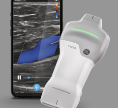
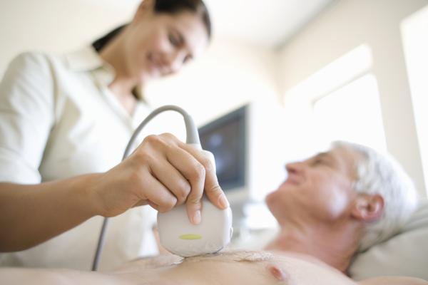
Whether in the ED or elsewhere in the hospital setting, patients experiencing cardiac arrest need swift and accurate resuscitation efforts. In addition to highly trained clinical teams, cardiac arrest patients can benefit from advances in point-of-care ultrasound (POCUS) technology.
According to the American College of Emergency Physician guidelines, transesophageal echocardiogram (TEE) allows physicians to maintain the standard of an ultrasound-informed resuscitation in the scenario of cardiac arrest, where transthoracic echocardiogram (TTE) is significantly limited.
Point-of-care transesophageal echocardiography (TEE) is becoming more widely used in cardiac arrest. According to experts, it has four main advantages over TTE: A reliable sonographic window, continuous imaging of the heart during resuscitation, elimination of pauses in chest compressions, and potential to detect abnormalities that are more difficult to visualize on TTE.
Simply put, with point-of-care TEE, patients can benefit from more accurate and efficient chest compressions while being evaluated with ultrasound — potentially making the response to a cardiac emergency more effective. Clinicians must act fast to ensure imaging does not interrupt or delay compressions, so having the right equipment and cardiac views can be critical.
No matter how seasoned an individual physician, cardiac arrest is a stressful situation requiring healthcare professionals to stay calm, composed and focused. Having the imaging tools on hand to address a resuscitation event gives physicians added confidence.
When choosing appropriate imaging equipment, providers should look for POCUS technology that offers exceptional image clarity on a large, adaptable clinical display for easy viewing by the entire clinical resuscitation team. Most importantly, the technology should be designed to provide real-time heart visualization during patient resuscitation. This feature helps resuscitation teams make rapid and potentially life-saving decisions at the bedside with the goal of improving shock and arrest outcomes for patients.
Choosing the right transducer for use during resuscitation is critical — it must allow clinicians to confidently assess patients during resuscitation and enable repeatable cardiac imaging regardless of patient condition or body habitus. It should also enable continuous image acquisitions both during compressions and during pulse checks, while not interfering with compressions or other procedures.
Ergonomic features are just as important, too. Transducers that are lightweight and offer easy manipulation while delivering exceptional image clarity can improve efficiency during a stressful cardiac emergency. One key vendor recently introduced its cardiac resuscitation exam type to address key needs of the clinical situation, leveraging insights from physician-leaders in POCUS.
Addressing Barriers to TEE, Improving Outcomes for IHCA
As mentioned earlier, in-hospital cardiac arrest can happen anywhere throughout the institution—in the ED, ICU, OR, or ward setting. However, barriers still exist to the widespread implementation of TEE in the ED and beyond. There is a continued need for training. The cost of purchasing, storing and reprocessing equipment is another concern for many healthcare organizations.
As one academic article recently noted, not all clinical environments have enough staff available to have a dedicated provider perform the ultrasound during pulse checks of a resuscitation. The authors note that artificial intelligence might play a greater role in the process in the future. As stated, if machine learning algorithms could identify the presence or absence of cardiac activity, it would significantly reduce the cognitive load on a single provider leading a resuscitation.
As a proven lifesaving procedure, both healthcare providers and vendors need to continue to take steps to address any barriers to the widespread implementation of TEE.
Today, even at the nation’s best hospitals where a code blue team is at the ready, many people who experience a sudden cardiac arrest are unlikely to survive. Reversing the high mortality rate associated with IHCA will require continued research, education and training, and technological innovation.
Safe and effective resuscitation depends first and foremost on competent and well-trained care teams, followed by the caliber of the tools they have available. Ultrasound provides tremendous information to aid in decision making during cardiac arrest. POCUS technology designed to support clinicians during the resuscitation process can be a life-saving tool.

Paul M. Bojarski is Senior Manager Market Development: Acute Care for Fujifilm Sonosite, Inc.
SIDEBAR
Cardiac Arrest in America: The Vitals
In the United States, sudden cardiac arrest is the leading cause of natural death. Both out-of-hospital cardiac arrest (OHCA) and in-hospital cardiac arrest (IHCA) are associated with high mortality rates. The vital statistics follow.
OHCA Facts:*
- Over 356,000 cases annually
- 90% fatal
- Location of OHCA in 2020: Residence (73.9%), public settings (15.1%), nursing homes (10.9%)
- Laypeople initiated CPR in 40.8% of OCHAs in 2020
IHCA Facts:**
- Over 290,000 cases annually
- 58% male
- Mean age: 66
- Point-of-care transesophageal echocardiography (TEE), recommended by the American College of Emergency Physicians, is becoming more widely used in resuscitation during IHCA
*Sudden Cardiac Arrest Foundation
** “In-Hospital Cardiac Arrest,” NIH/National Library of Medicine
https://www.ncbi.nlm.nih.gov/pmc/articles/PMC6482460/
Related ultrasound content:

 May 19, 2026