April 6, 2024 — Patients with heart failure who had a small shunt inserted between the heart’s left and right atria did not see any significant benefits overall compared with those who received a ...
Transesophageal Echo (TEE)
This channel includes news and new technology innovations for transesophageal echocardiography (TEE) ultrasound imaging used for diagnostics and procedural guidance. TEE is an invasive form cardiac ultrasound (echo), where the ultrasound probe is placed down the patient's throat, into the esophagus to allow for more detailed, clearer imaging of the heart than is available using typical transthoracic echo imaging. TEE is used extensively for transcatheter structural heart procedure guidance because of its ability to show soft tissue that is not visible on the angiography systems typically used in the cath lab.
-

-

Invented in 1896 by Enrico Salvioni, the fluoroscope remains a flagship technology of modern medicine. The live video X-ray image it provides can guide a catheter safely through a living patient’s ...
-

January 17, 2022 – As the increasing number of structural heart interventions are assisted by real-time imaging guidance, interventional echocardiography is being recognized as a subspecialty ...
-

June 4, 2020 — Intra-operative transesophageal echocardiography (TEE) is a versatile diagnostic and monitoring tool used to assist in patient management in a wide-range of cardiac surgical procedures ...

June 13, 2024 — Visura Technologies, Inc., a privately owned medical device company, has announced the TEECADE mini, an ...
 June 13, 2024
An Auxiliary CHD Diagnostic System Based on Multi-view and Multi-modal Transthoracic Echocardiograms
May 29, 2024 — Congenital heart disease (CHD) is one of the most common congenital anomalies worldwide, which brings a ...
May 29, 2024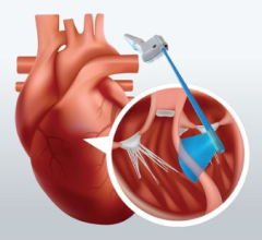
April 29, 2024 — FUJIFILM Healthcare Americas Corporation, a leading provider of diagnostic and enterprise imaging ...
April 29, 2024
April 6, 2024 — Patients with heart failure who had a small shunt inserted between the heart’s left and right atria did ...
April 06, 2024
January 31, 2024 — Royal Philips, a global leader in health technology, announced that its latest TEE transducer ...
January 31, 2024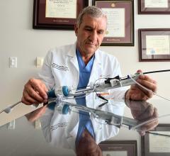
October 19, 2023 — The Dib UltraNav Transseptal Catheter System, which houses a needle and ultrasound in one system for ...
October 19, 2023
October 12, 2023 — Franklin Mountain Medical announced today that the Dib UltraNav Transseptal Catheter System, which ...
October 12, 2023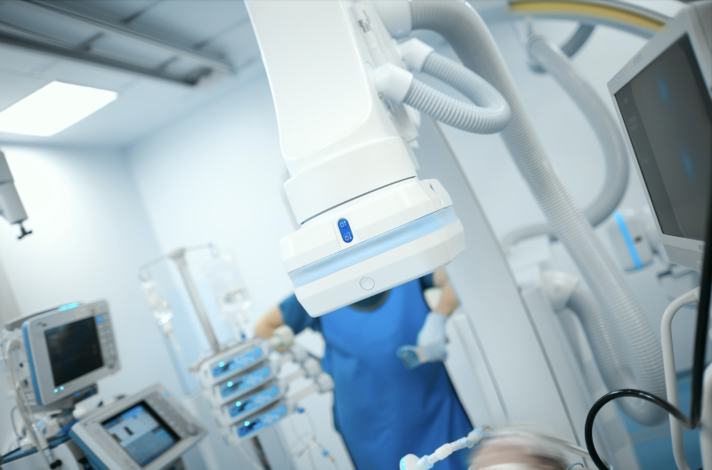
Feature | Cardiac Imaging | By Mohammad Sahebjalal, MD
Invented in 1896 by Enrico Salvioni, the fluoroscope remains a flagship technology of modern medicine. The live video X ...
May 04, 2023
March 28, 2023 — The Society for Cardiovascular Angiography and Interventions (SCAI) and the Heart Rhythm Society (HRS) ...
March 28, 2023
March 15, 2023 — Researchers at the University of Alabama at Birmingham Marnix E. Heersink School of Medicine published ...
March 15, 2023
Feature | Cardiovascular Ultrasound | By Paul M Bojarski
Sudden cardiac arrest is a complex and challenging medical emergency because it can occur anywhere at any time and is ...
August 23, 2022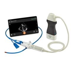
June 30, 2022 — Clarius Mobile Health, a leading provider of high-definition wireless ultrasound systems, and ImaCor Inc ...
June 30, 2022
June 8, 2022 — Us2.ai, a Singapore-based medtech firm backed by, IHH Healthcare, Heal Partners, Sequoia India, EDBI ...
June 08, 2022
June 7, 2022 — Visura Technologies, Inc., a privately-held medical device company dedicated to delivering state-of-the ...
June 07, 2022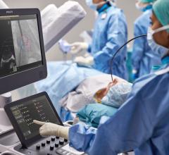 May 16, 2022