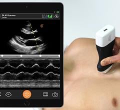
November 30, 2020 — The Clarius PA HD hand-held, wireless ultrasound scanner is now available for high resolution ...
Cardiac Imaging
The cardiac imaging channel includes the modalities of computed tomography (CT), cardiac ultrasound (echocardiography), magnetic resonance imaging (MRI), nuclear imaging (PET and SPECT), and angiography.
 November 30, 2020
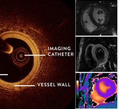
November 17, 2020 — Diagnostic imaging techniques were able to find the underlying cause of heart attack in many women ...
November 17, 2020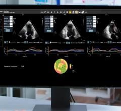
November 12, 2020 – Konica Minolta Healthcare Americas Inc. and DiA Imaging Analysis Ltd. jointly announce the expanded ...
November 12, 2020Sponsored Content

Feature
SPONSORED CONTENT — Studycast is a comprehensive imaging workflow system that allows healthcare professionals to work ...
June 17, 2025
Keith Ellis, M.D., is the director of cardiovascular services and the director of the Chest Pain Center at Houston ...
November 10, 2020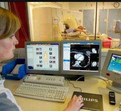
November 6, 2020 — The Society of Cardiovascular Computed Tomography (SCCT) has released a new training guideline, Train ...
November 09, 2020Feature | Radiation Dose Management | Dave Fornell, Editor
More than a decade ago, there was an alarming, rapid rise in ionizing radiation exposure in the U.S. population that was ...
November 04, 2020Sponsored Content

Feature | Cardiac Imaging
Cardiac positron emission tomography (PET) is growing in popularity among cardiologists because it provides the ability ...
March 05, 2024
October 29, 2020 — Contrast agents used to improve views of the heart on magnetic resonance imaging (MRI) carry a very ...
October 29, 2020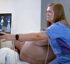
October 28, 2020 — Northwestern Memorial Hospital is the first hospital in the United States to purchase Caption Health ...
October 28, 2020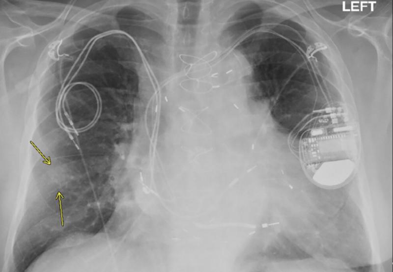
October 27, 2020 – Magnetic resonance imaging (MRI) examinations can be safely performed in patients with non-MR ...
October 27, 2020Sponsored Content

Blog | Enterprise Imaging
As medical advancements continue to push the boundaries of what is possible in the field of structural heart ...
August 10, 2023
News | FFR Technologies | Dave Fornell, Editor
October 22, 2020 – In the FORECAST randomized clinical trial, the use of fractional flow reserve (FFR) derived from ...
October 22, 2020
October 13, 2020 — GE Healthcare announced U.S. FDA 510k clearance for its Ultra Edition package on Vivid cardiovascular ...
October 15, 2020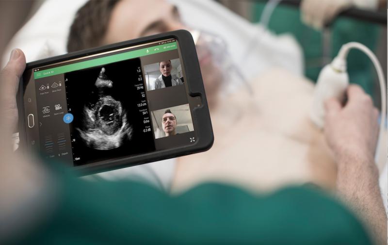
Feature | Point-of-Care Ultrasound (POCUS) | Alan Stoddart, Signify Research
For all the changes in medicine there are some things that seem resolutely stable. Chief among these is the idea that ...
October 08, 2020Sponsored Content

Feature | Cardiac Imaging
Discover the key features of cardiovascular structured reporting that drive adoption, including automated data flow, EHR ...
November 07, 2022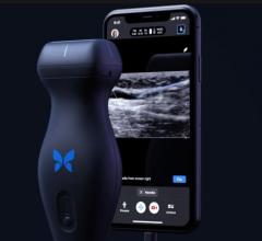
October 8, 2020 – Butterfly Network Inc. is launching its next-generation Butterfly iQ+ point-of-care-ultrasound (POCUS) ...
October 08, 2020
September 30, 2020 — Siemens Healthineers has introduced a new version of its c.cam dedicated cardiac nuclear medicine ...
October 07, 2020
October 2, 2020 — The new cardiac hybrid operating room (OR) at London Health Sciences Centre (LHSC) in London, Ontario ...
October 02, 2020