
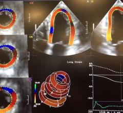
Marielle Scherrer-Crosbie, M.D., Ph.D., director of echocardiography at the Hospital of the University of Pennsylvania ...
Cardiac Imaging
The cardiac imaging channel includes the modalities of computed tomography (CT), cardiac ultrasound (echocardiography), magnetic resonance imaging (MRI), nuclear imaging (PET and SPECT), and angiography.

Judy Hung, M.D., director of echocardiography, Division of Cardiology, Massachusetts General Hospital, Boston, explains ...
 June 27, 2019
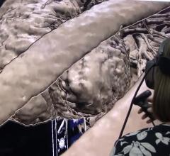
Roberto Lang, M.D., director of cardiac imaging at the University of Chicago, has been working with TomTec for the past ...
June 27, 2019Sponsored Content

Feature | Cardiac Imaging
Sponsored Content — According to the American Heart Association, cardiovascular disease is the leading cause of death in ...
March 13, 2026
Thomas Porter, M.D., FASE, the Theordore F. Hubbard Distinguished Chair of Cardiology and a professor of medicine at the ...
June 26, 2019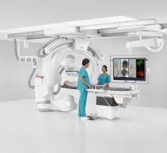
June 24, 2019 — Siemens Healthineers and Mentice AB announced the collaboration to fully integrate Mentice’s VIST ...
June 24, 2019
John Rumsfeld, M.D., Ph.D., FACC, American College Cardiology (ACC) chief innovation officer, and professor of medicine ...
June 20, 2019Sponsored Content
Feature
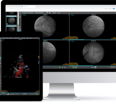
SPONSORED CONTENT — Studycast is a comprehensive imaging workflow system that allows healthcare professionals to work ...
June 17, 2025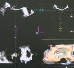
June 20, 2019 — Bay Labs announced that new data on the company’s first-of-its-kind deep learning investigational ...
June 20, 2019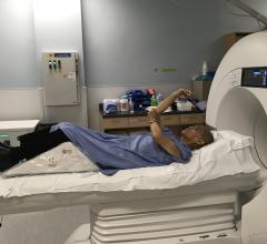
June 20, 2019 — A new type of care for cardiac patients at St. Paul’s Hospital, Vancouver, has the potential to reduce h ...
June 20, 2019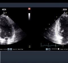
June 19, 2019 — DiA Imaging Analysis announced the presentation of two studies assessing the performance and accuracy of ...
June 19, 2019Sponsored Content

Feature | Cardiac Imaging
Cardiac positron emission tomography (PET) is growing in popularity among cardiologists because it provides the ability ...
March 05, 2024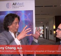
Anthony Chang, M.D., chief artificial intelligence officer, Children’s Hospital of Orange County (CHOC), and founder, AI ...
June 18, 2019
June 14, 2019 – A late-breaking study examined the effects of intravascular ultrasound (IVUS) guided drug-eluting stent ...
June 14, 2019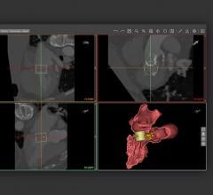
June 13, 2019 — Three-dimensional (3-D) printing software and solutions company Materialise has received U.S. Food and ...
June 13, 2019Sponsored Content
Blog | Enterprise Imaging
As medical advancements continue to push the boundaries of what is possible in the field of structural heart ...
August 10, 2023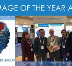
June 7, 2019 — The Amsterdam University Medical Center has won MR Solutions’ Image of the Year 2019 award for the best ...
June 07, 2019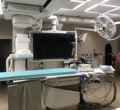
A discussion with Nicholas Bevins, Ph.D., vice chair, physics and research, and Jessica Harrington, RCIS. They explain ...
June 06, 2019
June 5, 2019 — The Society of Cardiovascular Computed Tomography (SCCT) will present the 2019 Gold Medal Award to Jonath ...
June 05, 2019