Figure 1: Segmentation performed on the left atrium, left ventricle and mitral bioprosthesis. Landmarks are placed on the site of optimal transseptal access into the left atrium and the mitral PVL.
Transcatheter interventions have become a mainstay in cardiac care as treatments for structural and congenital heart disease transition from open-heart surgery to minimally invasive procedures. These procedures, while minimally invasive for patients, often have added degrees of patient-related and procedural-related complexity.
“In order to make these complex procedures possible, we rely on multimodality cardiac imaging to improve what we as operators can visualize, both for planning and guidance” said Chad Kliger, M.D., M.S., director of structural heart disease at Lenox Hill Hospital in New York City.
Lenox Hill Heart and Lung, part of Lenox Hill Hospital, now uses the Artis pheno system from Siemens Healthineers. This advanced robotic C-arm fluoroscopy system allows for pre-procedural planning and intra-procedural imaging guidance during minimally invasive structural heart interventions.
Established in 1857, Lenox Hill Hospital, a member of Northwell Health, has a history of embracing cutting-edge technology to deliver high-quality care. The 652-bed facility has performed groundbreaking procedures that include the first angiocardiography and coronary angioplasty in the nation and the first placement of a drug-coated coronary stent.
A Changing Landscape in Complex Cardiac Cath Lab Imaging
Fluoroscopy has long been the cornerstone of interventional procedures thanks to the excellent device visualization and real-time feedback it provides. However, there is growing interest in limiting radiation and contrast dye exposure. In addition, fluoroscopy provides poor characterization of non-radiopaque structures and only provides 2-D projections of important 3-D cardiac anatomy, lacking essential spatial resolution.
While there have been recent significant improvements in 3-D/4-D transesophageal echocardiography (TEE) and cardiac computed tomographic angiography (CTA), using multiple imaging techniques independently in the middle of a procedure can be cumbersome and requires the operator to mentally fuse images. Looking for a solution that minimizes the limitations of each imaging modality, Lenox Hill Hospital turned to Siemens Healthineers’ cardiac fusion imaging technology, CTA-fluoro fusion (syngo X) and TEE-fluoro fusion (TrueFusion).
Integrating Fusion Imaging in the Hybrid OR
In March 2017, the U.S. Food and Drug Administration (FDA) cleared the Siemens Artis pheno system for use in minimally invasive procedures. Shortly after, in October 2017, Lenox Hill Hospital opened its new cardiovascular and thoracic surgery hybrid operating room suite featuring the Siemens Artis pheno, becoming the first advanced robotic system on the East Coast. With this system, fluoroscopic images can now be taken at a variety of angles as dictated by the procedure. The Artis pheno system also uses ultra-low dose radiation for imaging and produces images at a higher resolution and higher speed, resulting in less radiation exposure for both patients and the operating room team.
In addition, new fusion imaging software overlays information from both TEE and CTA onto live fluoroscopy. While fusing CT images with fluoroscopy by syngo X has been available for some time, fusing TEE with fluoroscopy recently received FDA clearance in September 2017. TrueFusion takes live 3-D TEE from the Siemens Acuson SC2000 and overlays key information, such as anatomical landmarks or valve models from the eSie valve modeling tool. Ultimately, these fusion images provide operators with real-time 3-D context that fluoroscopy alone never provided.
The basic steps of fusion imaging include segmentation, landmarking, co-registration and image overlay. After initial imaging acquisition, a 3-D model of cardiac structures of interest is generated either automatically or manually. Next, landmarks are placed to identify the cardiac defect for planned intervention. The CTA and/or TEE is subsequently co-registered within the X-ray space by matching the 3-D position and scale of both imaging modalities in two views separated by at least 30 degrees. For syngo X, co-registration can be achieved using contrast aortography or by existent radiopaque makers such as prosthetic valves or calcification; for TrueFusion, machine learning-based probe detection registers the 3-D TEE field-of-view. Lastly, the CTA/TEE with landmarks are overlaid onto fluoroscopy with the landmarks moving in real-time with the C-arm.
Fusion Aids Mitral Paravalvular Leak Closure
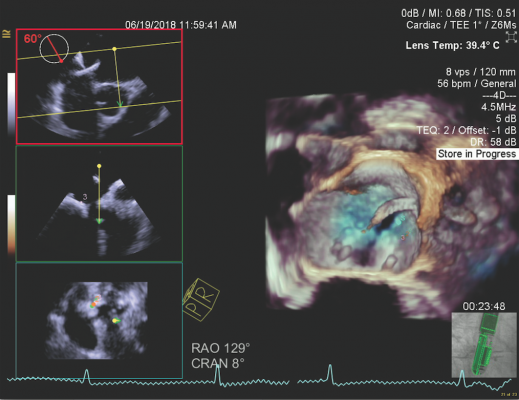
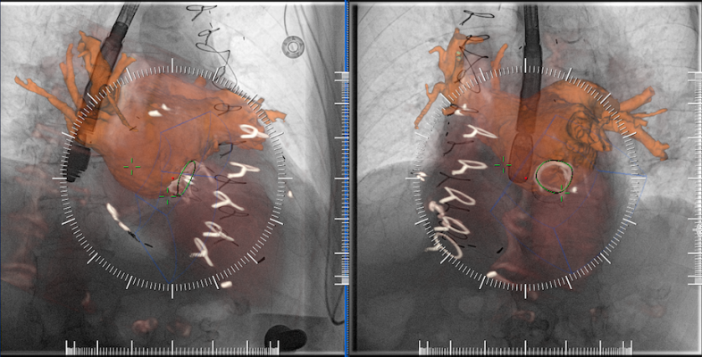
A mitral valve paravalvular leak (PVL) closure was one of the first procedures using syngo X and TrueFusion in the laboratory. An 80-year-old patient with multiple comorbidities had two previous mitral valve replacements with bioprosthetic valves, with the most recent in 2010. He presented with congestive heart failure (NYHA class III) and hemolytic anemia that required blood transfusions every two weeks. The pre-procedural CTA and intra-procedural TEE identified the PVL in the postero-medial location. (Figure 2)
In Figure 1, segmentation was performed of the left atrium, left ventricle and mitral bioprosthesis, and landmarks placed on the site of optimal transseptal access into the left atrium and the mitral PVL. In Figure 2, CTA and TEE images are co-registered in two views performed >30 degree apart. In Figures 3 and 4, TrueFusion and syngo X overlay can be seen. Successful percutaneous closure was performed using fusion imaging guidance with the placement of a single 10 mm Amplatzer Vascular Plug II; no residual regurgitation.
“Before using this software, I would have to split my focus between multiple individual screens showing a post-processed 3-D/4-D CTA, live TEE, fluoroscopy and hemodynamics. Now that information can be overlaid onto a single screen, I no longer have to mentally merge these different modalities and can focus more to the task at hand, especially in cases where navigating wires, catheters and devices are a challenge,” Kliger said following completion of the mitral PVL procedure.
Providing Operational Benefits in the Cath Lab
Overlaying TEE and CTA information simultaneously on live fluoroscopy imaging can provide additional helpful information during percutaneous procedures. With the Siemens fusion imaging suite, Lenox Hill Hospital has a unique solution that can drastically change how minimally invasive procedures are performed. Using this technology, Lenox Hill Heart and Lung continues to provide high-quality care to its structural heart patients, while potentially reducing procedure time and decreasing contrast dye and radiation delivered.
For more information: usa.healthcare.siemens.com


 May 29, 2026