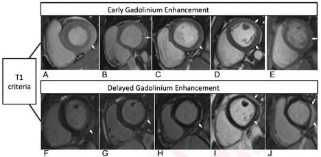
T1-Based Criteria for Myocarditis on Cardiac MRI in Patients With Recent COVID-19 mRNA Vaccination.[1] Early gadolinium enhancement (EGE) compared with precontrast SSFP sequence (not shown) is observed on early post-contrast short-axis SSFP images in (A) 16-year-old male, (B) 17-year-old male, (C) 16-year-old male, and (D) 19-year-old male, and on early postcontrast short-axis perfusion image in (E) 17-year-old male (arrow, A-E). Late gadolinium enhancement (LGE) is also present (arrows) in all 5 patients (F, G, H, I, and J; same patients as in A, B, C, D, and E, respectively). EGE and LGE predominantly affect subepicardium of the inferior, inferolateral, or anterolateral walls. EGE extends beyond confines of LGE in patient shown in (A) and (F) and in (B) and (G). Basilar LV involvement is present in patient shown in (A) and (F), (B) and (G), and (C) and (H). Mid-cavity LV involvement is present in patient shown in (D) and (I) and in (E) and (J).
November 1, 2021 — According to ARRS’ American Journal of Roentgenology (AJR), radiologists need to be cognizant of the association between coronavirus disease (COVID-19) mRNA vaccination and myocarditis, as well as the role of cardiac MRI for assessing suspected myocarditis post-vaccination.[1]
“In this small case series, all patients with myocarditis after COVID-19 vaccination were adolescent males and had a favorable initial clinical course,” explained first author Lydia Chelala from University of Chicago Medicine. Noting that every patient’s cardiac MRI examination showed findings typical of myocarditis from other causes, “late gadolinium enhancement (LGE) persisted in two patients undergoing repeat MRI.”
Chelala and team’s retrospective study included patients who underwent cardiac MRI between May 14, 2021 and June 14, 2021 for suspected myocarditis within 2 weeks of COVID-19 mRNA vaccination — without known prior COVID-19. With clinical presentation, hospital course, and post-discharge events recorded, the cardiac MRI examinations were reviewed in consensus by a cardiothoracic radiologist and cardiothoracic imaging fellow.
Of the 52 patients who underwent cardiac MRI during the study period, Chelala and colleagues identified 5 male patients (age range, 16–19 years; mean age, 17.2 years) who presented within 4 days of the second dose of COVID-19 mRNA vaccine. After mean hospitalization length of 4.8 days, all 5 patients were discharged in stable condition with improved or resolved symptoms. However, two patients underwent repeat cardiac MRI that showed persistent, albeit decreased, LGE.
Acknowledging that their article is the first report to describe additional results of short-term follow-up in this patient population, the authors of this AJR article also conceded, “the observations do not establish causality.”
For more information: www.arrs.org
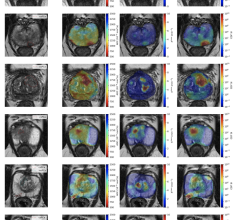
![Figure 2. Qualitative and quantitative T2-based criteria for myocarditis on cardiac MRI in patients with recent COVID-19. mRNA vaccination. Qualitative criterion was focal myocardial edema and is shown on 4-chamber precontrast SSFP image (arrow) in 16-year-old male (A) and 17-year-old male (B). Quantitative criteria included T2 parametric mapping and quantification of myocardial signal intensity ratios. Focal subepicardial edema of the basilar left ventricular inferior wall (arrow) is shown on source image from T2 mapping in 17-year-old male (C). Increased myocardial signal intensity ratio of 3.0 at mid-cavity is shown in 19-year-old male (D). Qualitative focal myocardial edema is also shown on short-axis precontrast SSFP image (arrow) in 17-year-old male (E). Patients in A-E correspond with patients A-E in Table 1, respectively.[1]](/sites/default/files/imce_images/MRI_COVID_vaccine_Myocarditis_AJR2.jpg)
Figure 2. Qualitative and quantitative T2-based criteria for myocarditis on cardiac MRI in patients with recent COVID-19. mRNA vaccination. Qualitative criterion was focal myocardial edema and is shown on 4-chamber precontrast SSFP image (arrow) in 16-year-old male (A) and 17-year-old male (B). Quantitative criteria included T2 parametric mapping and quantification of myocardial signal intensity ratios. Focal subepicardial edema of the basilar left ventricular inferior wall (arrow) is shown on source image from T2 mapping in 17-year-old male (C). Increased myocardial signal intensity ratio of 3.0 at mid-cavity is shown in 19-year-old male (D). Qualitative focal myocardial edema is also shown on short-axis precontrast SSFP image (arrow) in 17-year-old male (E). Patients in A-E correspond with patients A-E in Table 1, respectively.[1]
COVID Vaccine Causes Myocarditis in Small Number of Younger Males
A small number of patients develop inflammation leading to myocarditis after receiving the messenger RNA (mRNA) COVID-19 vaccines from Pfizer and Moderna. This raised concerns earlier in 2021 after numbers for this adverse reaction to the vaccine jumped in April and May and vaccinations were opened up to all people ages 12 and older. The U.S. Food and Drug Administration (FDA) added a warning on the Moderna and Pfizer mRNA vaccine fact sheets about myocarditis being a rare, but possible side effect.
The agency also said the side effect is very rare and patients generally recover quickly, The CDC said the risk of COVID is high and potentially more serious, so the CDC is continuing to encourage people to get the vaccine.
The patients effected are mostly men aged 16-19, and symptoms usually occurred after the second dose, explained Tom Shimabukuro, M.D., MPH, MBA, deputy director of the CDC Immunization Safety Office, and a member of the Vaccine Safety Team, CDC COVID-19 Vaccine Task Force. He said most of the patients were hospitalized, but 95 percent of the cases were considered mild. While there were cases of myocarditis in all age groups between 12-50, the majority of cases are concentrated in the 16-28 age group.
With the vaccine now opening up to younger age groups, this is a possible side effect the FDA is warning clinicians to watch for.
Read more:
FDA Adds Myocarditis Warning to COVID mRNA Vaccine Clinician Fact Sheets
Small Number of Patients Have Myocarditis-like Illness After COVID-19 Vaccination
Overview of Myocarditis Cases Caused by the COVID-19 Vaccine
Related COVID-19 Content:
Medical AI Models Rely on 'Shortcuts' That Could Lead to Misdiagnosis of COVID-19
CT Provides Best Diagnosis for Novel Coronavirus (COVID-19)
SNMMI Image of the Year: PET Imaging Measures Cognitive Impairment in COVID-19 Patients
Cardiac MRI Effective in Detecting Asymptomatic, Symptomatic Myocarditis in Athletes
PHOTO GALLERY: How COVID-19 Appears on Medical Imaging
VIDEO: How to Image COVID-19 and Radiological Presentations of the Virus — Interview with Margarita Revzin, M.D.
How Does COVID-19 Appear in the Lungs?
Find more radiology related COVID news and video
Reference:

 April 27, 2026