January 20, 2017 — A new document from the European Association of Cardiovascular Imaging (EACVI) and the American Society of Echocardiography (ASE) provides a comprehensive review of the optimal application of 3-D echocardiography for patients with congenital heart disease (CHD).
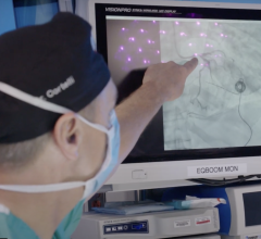
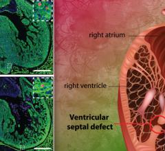
Congenital heart disease (CHD) is the most common form of birth defect worldwide, affecting 40,000 children born annually in the United States alone. Standard two-dimensional echocardiography, ultrasound of the heart and vascular system, has always been an essential non-invasive diagnostic tool for pediatric cardiologists, but recent advances in technology have led to the increasing importance of three-dimensional echocardiography in this field.
The paper, “Three-dimensional Echocardiography in Congenital Heart Disease: An Expert Consensus Document from the European Association of Cardiovascular Imaging and the American Society of Echocardiography,” will appear in the January issue of the Journal of the American Society of Echocardiography (JASE), but is available now online.
ASE’s co-chair of the writing group, Girish S. Shirali, MBBS, FASE of Children’s Mercy Hospital in Kansas City, Mo., commented, “Pediatric cardiology is unique in the wide range of structural abnormalities that we encounter, and for which echocardiography is the most common — and frequently the only — preoperative diagnostic modality that is used. In the absence of clear guidelines for its use, pediatric cardiac centers from a wide range of geographies have developed a wide range of approaches to 3-D echocardiography. This document helps establish a common logic and structure for the display of images, and uses the collective experience of a large group of echocardiographers and sonographers to help develop a common language and approach to 3-D echocardiography. We believe this modality is a powerful addition to the pediatric echocardiographer's armamentarium, and believe that the current offering will go a long way in enhancing the adoption of 3-D echocardiography for our patients' benefit.”
The document outlines in detail the technical considerations and imaging techniques, as well as the value that 3-D echocardiography can add to the management of specific congenital heart defects. It also details the use of 3-D echocardiography to guide catheter-based interventions, which have become increasingly used in recent years, and addresses the need for specific training and educational pathways specific to 3-D echocardiography. The document includes several useful tables summarizing techniques and advantages of 3-D echocardiography for specific types of CHD, as well as 17 figures to illustrate various concepts.
In conjunction with the publication of the guideline document, Shirali will conduct a live webinar, including a question and answer section, on Feb. 15, 2017 at 5:00 PM Eastern Time. The webinar will be available for free to all ASE members and open to all other clinicians for just $25; registration is available at ASEUniversity.org/ase/live/9. This webinar, and all ASE-hosted guideline webinars, are available on ASEUniversity.org to facilitate education for those who cannot attend the live webcast.
The full guideline document is available on the Journal of American Society of Echocardiography (JASE) website (OnlineJASE.com). This document and all ASE Guideline documents are also available to the medical community at ASEcho.org/Guidelines.
For more information: www.asecho.org

 March 31, 2025