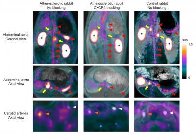
Note the high uptake of Ga-68-pentixafor on multi-planar reconstructions in the organs expressing CXCR4 such as the spleen (red arrows) and adrenal glands (yellow arrows), which was nearly completely blocked by the pre-injection of AMD 3100, a potent CXCR4 inhibitor. Strong accumulation of Ga-68-pentixafor was also found in the kidneys (asterisks) reflecting the renal clearance of the tracer. In addition, high, focal activities were detected in the abdominal aorta (red arrowheads) and right carotid artery (orange arrowheads) of atherosclerotic rabbits, whereas no significant signal could be detected in the non-injured left carotid artery (white arrowheads) of atherosclerotic and control rabbits, as well as in the abdominal aorta and right carotid artery of control rabbits. Furthermore, focal activities detected with PET in atherosclerotic plaques of the abdominal aorta and the right carotid artery decreased significantly when the same rabbit was re-imaged after blocking CXCR4 receptors. Image courtesy of Fabien Hyafil, M.D., Ph.D., Department of Nuclear Medicine, Klinikum Rechts der Isar, Technische Universität München, Munich, Germany
March 3, 2017 — In the featured article of the March 2017 issue of The Journal of Nuclear Medicine, researchers demonstrate that a new positron emission tomography (PET) radiotracer, gallium-68 (Ga-68)-pentixafor, can quickly and non-invasively identify life-threatening atherosclerotic plaques. The tracer binds to the CXCR4 receptor on inflammatory cells present in atherosclerotic plaques — making it possible to find and treat atherosclerosis early.
Atherosclerosis represents the main cause of heart attack and stroke. According to the Centers for Disease Control and Prevention, every year about 735,000 Americans have a heart attack and 800,000 have a stroke. Stroke kills more than 130,000 Americans a year, and about 610,000 die from cardiovascular disease.
Atherosclerosis develops over decades with the progressive accumulation of lipids, inflammatory cells and connective tissue within the inner layer of arterial walls leading to a local thickening of the vascular wall called atherosclerotic plaque. These plaques can remain asymptomatic for years, but an inflammatory reaction can develop causing the plaques to rupture and stimulate clot formation. If a clot completely blocks an artery, no oxygen can reach the downstream tissue, resulting in the sudden development of heart attack or stroke. The challenge is to identify patients with these dangerous atherosclerotic plaques before a heart attack or stroke occurs.
Currently, there is no clinically available non-invasive imaging technique specifically to detect inflammation. F-18-fluorodeoxyglucose (FDG)-PET is being used but has important limitations. It is taken up by many cells other than inflammatory cells, including cardiac and brain cells. The strong signal present in the organs next to the arteries limits the precise analysis of the radiotracer uptake in atherosclerotic plaques. In addition, patients need to fast at least six hours before FDG injection to avoid interferences with blood sugar and muscular uptake of the tracer that impair image quality.
"Ga-68-pentixafor binds more specifically to inflammatory cells than FDG and does not require the patient to fast for six hours before imaging," explained Fabien Hyafil, M.D., Ph.D., of Klinikum Rechts der Isar, Munich, Germany, and Bichat University Hospital, Assistance Publique, Hôpitaux de Paris, Paris, France.
In the study, the specific binding of Ga-68-pentixafor to inflammatory cells located in atherosclerotic plaques was first validated in an animal model. Seven atherosclerotic rabbits and five controls were imaged on a PET-MRI (magnetic resonance imaging) system after injection of the tracer. Resulting images clearly showed inflammation in plaques in the abdominal aorta and right carotid artery of the atherosclerotic rabbits. The researchers also confirmed with a small number of human patients that the radiotracer detected atherosclerotic plaques located in their carotid arteries.
Hyafil emphasized, "This new radiotracer will strongly facilitate the imaging of inflammation in atherosclerotic plaques with PET and hopefully support the early detection and treatment of atherosclerosis, thus preventing heart attack or stroke."
This work was supported by the European Research Council Executive Agency through a Multimodal Molecular Imaging Advanced Research Grant (Grant number 294582), the Deutsche Forschungsgemeinschaft (SFB 824-B5 and SFB 1123-A1), and Deutsches Zentrum für Herz-Kreislauf Forschung through a high-risk, high-volume grant.
For more information: www.jnm.snmjournals.org
References
Hyafil, F., Pelisek, J., Laitinen, I., Schottelius, M., et al. "Imaging the Cytokine Receptor CXCR4 in Atherosclerotic Plaques with the Radiotracer Ga-68-Pentixafor for PET," Journal of Nuclear Medicine. Published online Oct. 27, 2016. DOI: 10.2967/jnumed.116.179663
Read the article "Recent Advances in Cardiac Nuclear Imaging Technology."
Watch the VIDEO "PET vs. SPECT in Nuclear Cardiology and Recent Advances in Technology." An interview with Prem Soman, M.D., director of nuclear cardiology at the Heart and Vascular Institute, University of Pittsburgh, and president-elect of the American Society of Nuclear Cardiology (ASNC), explained advances in PET and SPECT imaging.
Watch the VIDEO "Trends in Nuclear Cardiology Imaging." A discussion with David Wolinsky, M.D., director of nuclear cardiology at Cleveland Clinic Florida and past-president of the American Society of Nuclear Cardiology (ASNC), discusses advancements in nuclear imaging and some of the issues facing the subspecialty.

 November 12, 2025