News | Cardiovascular Information Systems (CVIS) | October 17, 2019

USF Health Expands Digisonics System With Vascular Reporting

Vascular reporting module will fully integrate with Epic EMR for full documentation in the USF Health Vascular Surgery Department

USF Health Expands Digisonics System With Vascular Reporting

October 17, 2019 — University of South Florida (USF) Health in Tampa, Fla., has enhanced their use of the Digisonics Cardiovascular Information System to include vascular reporting for improved workflow automation and efficiency.

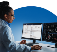
The Digisonics vascular reporting module, which is compliant with Intersocietal Accreditation Commission (IAC) standards and guidelines, will enable the Vascular Surgery Department to quickly document velocities, flow, pressures, clinical indications, findings and general conclusions. Seamless integration with the facility’s doppler ultrasound systems will automate transmission of patient information and measurement data directly into the report, translating to improved turnaround times and accuracy by eliminating redundant data entry workflows.

User-friendly drawing tools allow clinicians to quickly modify built-in vascular diagrams which can be included on the report, depicting a comprehensive view of the patient’s anatomy and disease. Vascular images can be quickly viewed at the workstation via a slideshow mode as well as embedded directly in the report with annotation capabilities.

The Digisonics solution will be fully integrated with the facility’s Epic electronic medical record (EMR) for improved interoperability and ease of use. Clinicians will have the convenience of immediate access to the finalized formatted report (with embedded images and diagrams) and study images directly from a link in the Epic patient record, connected to WebView, Digisonics’ universal viewer application.  

As a result of expanding Digisonics for vascular, USF Health has standardized its reporting across departments for the best quality of patient care.

For more information: www.digisonics.com


Related Content

Feature | Cardiac Imaging | By Mohammad Sahebjalal, MD

Invented in 1896 by Enrico Salvioni, the fluoroscope remains a flagship technology of modern medicine. The live video X ...

Home May 04, 2023
Home
News | Cardiac Imaging

April 26, 2023 — In the majority of cases, graft failure after heart transplantation is attributable to abnormalities ...

Home April 26, 2023
Home
News | Cardiac Imaging

April 14, 2023 — Genetesis, Inc., the leader in biomagnetic cardiac imaging solutions, today announced that the U.S ...

Home April 14, 2023
Home
News | Cardiac Imaging

February 8, 2023 — Carestream Health is partnering with Robarts Research Institute to increase and demonstrate the ...

Home February 08, 2023
Home
Videos | Cardiac Imaging

Artificial intelligence and general consolidation were two top cardiology trends at RSNA22. ITN/DAIC spoke with Val ...

Home January 23, 2023
Home
News | Cardiac Imaging

January 13, 2023 — Nuclear stress testing performed with single-photon emission computed tomography (SPECT) is the most ...

Home January 13, 2023
Home
News | Cardiac Imaging

November 29, 2022 — Researchers have developed a deep learning model that uses a single chest X-ray to predict the 10 ...

Home November 29, 2022
Home
News | Cardiac Imaging

November 28, 2022 – UltraSight, an Israeli-based digital health pioneer transforming cardiac imaging through the power ...

Home November 28, 2022
Home
News | Cardiac Imaging

November 28, 2022 — Konica Minolta Healthcare Americas, Inc., a leader in medical diagnostic imaging and healthcare ...

Home November 28, 2022
Home
Feature | Cardiac Imaging

Discover the key features of cardiovascular structured reporting that drive adoption, including automated data flow, EHR ...

Home November 07, 2022
Home
Subscribe Now