The Vivid 7 Dimension is an extraordinary cardiovascular ultrasound system built on a robust platform that empowers medical practitioners to utilize a diverse set of quantitative tools for increasing clinical confidence.
GE’s real-time 4D imaging and real-time full volume imaging enable the user to acquire depictions of the entire heart, helping to increase diagnostic confidence. Real-time 4D color Doppler, and real-time full volume color Doppler imaging, offer instant display of 4D color for detailed visualization of hemodynamics, including the ability to quantify regurgitant lesions.
As outlined in this case, 4D imaging provides incremental value for assessing the mitral valve:
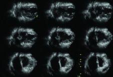
• 9 Slice enables the user to visualize regional wall motion in more detail by slicing the left ventricle of a 4D full-volume dataset into nine equidistant short-axis views.
• The 4D dataset can be further manipulated in order to interrogate specific areas of the mitral valve not visible with conventional 2D imaging.
• Full volume color Doppler imaging can be used to assess hemodynamic information in color and in real-time for further evaluation of valve function.
Clinical confidence can be increased using this comprehensive 4D imaging technology to generate an overall characterization of a patient’s valvular anatomy and pathology.
With this particular case at St. Francis Hospital, the need to complete a transesophageal exam was eliminated due to the quality of detailed images, and data generated using the less invasive transthoracic 4D imaging approach.
The patient is a 57-year-old female, with a history of mitral valve prolapse and mitral regurgitation, who was referred to the Echocardiography Laboratory at St. Francis Hospital for assessment of mitral regurgitation severity and suitability for repair.
Transthoracic two-dimensional echocardiography, using a Vivid 7 M4S matrix-array transducer from GE Healthcare, revealed myxomatous mitral disease and severe 4 mitral regurgitation. Proximal isovelocity surface area (PISA) calculations revealed an effective regurgitant orifice area (ERO) of 0.7 cm, and a calculated regurgitant volume of 150 ml/beat. However, the precise anatomic pathological details, segment involvement and consequent suitability for valve repair could not be ascertained.
Therefore, 4D transthoracic echocardiography was performed using a 3V matrix array transducer using the full-volume mode from the apical 4-chamber position. The full-volume 4D dataset was manipulated off-line utilizing an EchoPAC workstation. The 9-slice view was first inspected to exclude any stitching artifact.
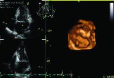
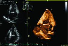
It was then rotated to visualize the myxomatous mitral leaflet in several slices, confirming the etiology of the mitral pathology. (Figure 1) The 4D dataset was further manipulated using the “angle” feature to visualize coronal sections of anterior and posterior leaflets. By “translating” the viewing plane anteriorly and posteriorly, a partially flail segment could be easily identified. (Figure 2)
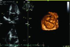
To further clarify the specific segments involved, the “angle” feature was again utilized to obtain axial sections of the mitral valve. “Box” and “crop” features were used to remove extraneous data surrounding the mitral valve. The viewing plane was then “translated” and aligned with the mitral annulus by “rotation” along the x and y-axes to obtain an enface view of the mitral valve from the left atrium.
The prolapse was found to involve P2 and P3 segments of the mitral valve with a clearly visualized flail segment. (Figure 3 and 4)
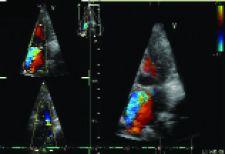
Since the anterior leaflet was spared, the valve was deemed suitable for repair by quadrangular resection. A full-volume 4D color flow acquisition was obtained, and manipulated to appreciate the complex and eccentric anteromedially-directed jet. (Figure 5)
Hence, a complete characterization of the patient’s valvular anatomy, pathology, severity and suitability was possible by utilizing transthoracic 2D and 4D data, obviating the need for transesophageal echocardiography.

 May 19, 2026