February 27, 2018 – A new study published in the Journal of the American College of Cardiology confirms non-invasive cardiac magnetic resonance (CMR) provides a highly accurate means of assessing mitral regurgitation in patients with valvular heart disease. Seth Uretsky, M.D., medical director of cardiovascular imaging and associate director of the Cardiovascular Fellowship Program at Atlantic Health System, served as the study’s lead author.
Uretsky presented his research findings at CMR 2018 in Barcelona, Spain, Jan. 31-Feb. 3. CMR 2018 was a joint meeting organized by the European Association of Cardiovascular Imaging (EACVI), a registered branch of the European Society of Cardiology, and the Society for Cardiovascular Magnetic Resonance (SCMR).
Mitral regurgitation is a common form of valvular heart disease affecting approximately 2 million Americans. It is a condition in which the heart's mitral valve does not close tightly, which allows blood to flow backward in the heart. Symptoms include shortness of breath, fatigue, lightheadedness and a rapid, fluttering heartbeat. Some people may not need treatment, however, more severe cases may require medication or surgery.
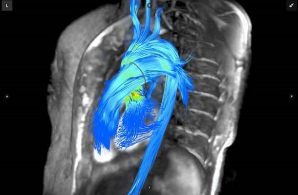
The current guidelines call for using echocardiography to determine the severity of mitral regurgitation. However, more physicians are using CMR without contrast to measure the volume of regurgitation. The use of CMR provides a highly accurate and reproducible assessment of the left and right chamber size and function, and has become the gold standard for evaluating cardiac chamber size, the review confirms. CMR complements echocardiography and, in some patients, is exceptionally helpful in determining how severe the leakage is.
“Clear, accurate imaging is critical to helping cardiologists accurately diagnose and confirm heart problems,” said Uretsky. “By using more cardiac magnetic resonance in patients with mitral regurgitation, cardiologists are able to better understand the extent of the lesion and determine the most appropriate treatment approach. For some patients, that may be the difference between having surgery to correct the valve or not.”
“Many patients don’t understand that cardiac imaging is not the same everywhere — for instance, a cardiac MRI performed at a center that doesn’t have updated technology, skilled technicians and an expert reading the images may provide an incomplete or inaccurate assessment of heart problems,” said Linda D. Gillam, M.D., MPH, FACC, the Dorothy and Lloyd Huck Chair of Cardiovascular Medicine at Morristown Medical Center/Atlantic Health System. “Patients should seek out a facility that provides high-quality imaging particularly if there are important management decisions to be made.”
The paper is available on the Journal of the American College of Cardiology website.
For more information: www.onlinejacc.org
References
Uretsky S., Argulian E., Narula J., Wolff S.D., et al. "Use of Cardiac Magnetic Resonance Imaging in Assessing Mitral Regurgitation: Current Evidence." Journal of the American College of Cardiology. doi: 10.1016/j.jacc.2017.12.009.

 June 12, 2026


