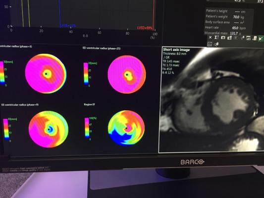
September 25, 2019 – Cardiac magnetic resonance imaging (MRI) analysis can be performed significantly faster with similar precision to experts when using automated machine learning, according to new research. The study was published in Circulation: Cardiovascular Imaging, an American Heart Association journal.[1]
Currently, analyzing heart function on cardiac MRI scans takes approximately 13 minutes for humans. Utilizing artificial intelligence (AI) in the form of machine learning, a scan can be analyzed with comparable precision in approximately four seconds.
Healthcare professionals regularly use cardiac MRI scans to make measurements of heart structure and function that guide patient care and treatment recommendations. Many important clinical decisions including timing of cardiac surgery, implantation of defibrillators, and continuing or stopping cardiotoxic chemotherapy, rely on accurate and precise measurements. Improving the performance of these measures could potentially improve patient management and outcomes.
In the U.K., where the study was conducted, it is estimated that more than 150,000 cardiac MRI scans are performed each year. Based on the number of scans per year, researchers believe that utilizing AI to read scans could potentially lead to saving 54 clinician-days per year at each U.K. health center.
Researchers trained a neural network to read the cardiac MRI scans and the results of almost 600 patients. When the AI was tested for precision compared to an expert and trainee on 110 separate patients from multiple centers, researchers found that there was no significant difference in accuracy.
“Cardiovascular MRI offers unparalleled image quality for assessing heart structure and function; however, current manual analysis remains basic and outdated. Automated machine learning techniques offer the potential to change this and radically improve efficiency, and we look forward to further research that could validate its superiority to human analysis,” said study author Charlotte Manisty, M.D. Ph.D. “Our dataset of patients with a range of heart diseases who received scans enabled us to demonstrate that the greatest sources of measurement error arise from human factors. This indicates that automated techniques are at least as good as humans, with the potential soon to be ‘super-human’ — transforming clinical and research measurement precision.”
Although the study did not demonstrate superiority of AI over human experts and was not used prospectively for clinical assessment of patient outcomes, this study highlights the potential that such techniques could have in the future to improve analysis and influence clinical decision making for patients with heart disease.
For more information: www.ahajournals.org/journal/circimaging
Reference
1. Bhuva A.N., Bai W., Lau C., et al. A Multicenter, Scan-Rescan, Human and Machine Learning CMR Study to Test Generalizability and Precision in Imaging Biomarker Analysis. Circulation: Cardiovascular Imaging, published online Sept. 24, 2019. https://doi.org/10.1161/CIRCIMAGING.119.009214

 May 20, 2026