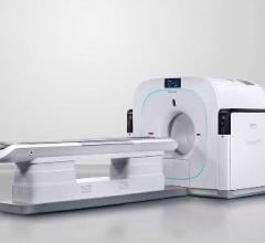
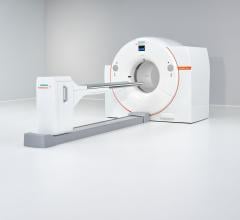
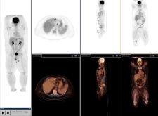
PET/CT in molecular imaging takes a 'photograph' of biological targets or pathways in the body for 'personalized medicine.' Photo courtesy of Siemens.
As in most partnerships, rarely are all things equal. Such is the case with hybrid PET/CT systems, where the vast majority of the improvements have benefited the CT side of the scanner, in particular with increased CT imaging speeds, according to Medhat M. Osman, M.D., Ph.D., at the School of Medicine at St. Louis University. This has left the medical community anxiously awaiting more substantial changes to the PET side of the PET/CT scanner.
Over the next 10 years, the hybrid PET/CT modality is expected to surpass dedicated PET devices in terms of adoption, as the combined functional and anatomical information produced by the hybrid has proven increasingly beneficial in oncology and cardiology. PET/CT will also play an important role in the future of molecular imaging, which aims to “photograph” biological targets or pathways in the body for “personalized medicine,” which will eventually provide patient-specific information that allows clinicians to tailor the treatment of disease. These developments have charged the leading PET/CT manufacturers, GE Healthcare, Siemens, Hitachi and Philips, with enhancing image quality in the hybrid – particularly in PET. What is intriguing is that each manufacturer is approaching this challenge in its own way.
With a growing focus on cardiology applications, GE Healthcare strives to enhance image quality through dynamic, gated PET and volumetric CT. The company introduced at the end of 2005 the Discovery VCT, a system that aims to convey a clearer view of the heart. The system combines PET’s dynamic and gating data, acquiring a comprehensive view of the heart and coronary arteries with submillimeter resolution in five seconds, with volumetric CT used to visualize the anatomy of the heart’s blood vessels. The VCT also features VUEPoint reconstruction, offering fully iterative reconstruction technology in 2-D and 3-D acquisition modes.
The angle that Siemens has carved out for improving PET/CT images is focusing on increased count rates. Siemens has adopted this approach by introducing TruePoint technology into its Biograph system. Designed for nuclear medicine applications where clinicians require extremely precise and detailed images to pinpoint minute lesions and arteries, True Point reportedly adds 33 percent more axial volume coverage to its PET/CT scanner, extending the field of view and improving counts by more than 78 percent.
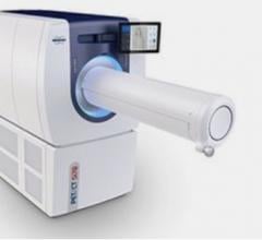
Hitachi’s SceptreP3 employs Dual Attenuation Correction (DAC) technology, which allows the combination of both CT and sealed source attenuation correction to effectively image patients with metal implants. In addition, the Non-Rigid Fusion 7-D algorithm provides precise registration by correcting for respiration differences between PET and CT acquisitions.
Time-of-Flight Takes Off
One technology used in PET imaging that has resurfaced in PET/CT systems is time-of-flight (TOF). The idea of TOF is not a new concept. TOF PET scanners were developed in the 1980s and at that time were used chiefly in research. Dr. Osman notes that TOF was a well-known academic concept from a physics aspect, and it was believed for many years that it would be a powerful addition to the PET modality. But due to various hurdles, it did not become a reality.
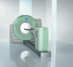
Last year, however, Philips Medical Systems introduced the Gemini TF PET/CT, which provides what Philips calls TruFlight, its own version of TOF technology. The Gemini TF is a high-performance, fully 3-dimensional, time-of-flight PET scanner combined with a Brilliance CT scanner (currently up to 64-slices). The PET scanner uses lutetiumyttrium oxyorthosilicate (LYSO) crystals that are placed in an Anger-logic detector, accomplishing a light spread in the detector that is uniform. The scanner was designed by Philips to be used as a high-performance conventional PET scanner in its own right, as well as a TOF scanner to provide improved timing resolution.
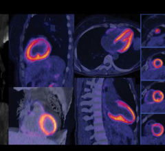
“TruFlight is a very sophisticated stopwatch,” said Dr. Osman. “Philips’ time-of-flight technology measures the actual time difference between the detection of two simultaneous and almost coincident gamma rays. This difference in detection times of the gamma rays observed by the PET scanner is used to more accurately localize the annihilation origin. It is the improvement in event localization that ultimately reduces image data noise, producing a higher quality image, allowing for lower tracer dose amounts in patients and shorter imaging times.”
TOF Better For Bariatrics?
TOF helps to reduce imaging noise, which is associated with acquiring images of obese patients. “The idea of noise and the resultant lower quality images seen when imaging obese patients is not being discussed enough,” noted Dr. Osman. He finds this especially troubling given that a large number of patients being scanned today fall into a body mass index range of overweight to obese. His concern is strengthened by numerous studies released in the past several years discussing America’s obesity problem.
The quality of PET images for obese patients obtained on non-time of flight scanners, according to Dr. Osman, are suboptimal. “The two options typically used to make up for suboptimal images, specifically injecting more radioactive tracer into the patient and utilizing higher imaging timeframes, do not adequately compensate for the suboptimal image quality. The time-of-flight technology alleviates this suboptimal image,” said Dr. Osman. In his experience, TOF images exhibit a much higher quality on obese patients than those images acquired not using the technology, with injected tracer amounts and image acquisition parameters remaining the same during both image acquisitions.
Since the introduction of the Gemini, Suleman Surti, Ph.D., research assistant professor at the University of Pennsylvania, has led a study to evaluate its intrinsic TOF capability and its impact on the reconstructed images. Dr. Surti and his fellow researchers “performed experiments with this scanner using reproducible phantoms mimicking the presence of lesions in a patient.” The measurement capability of the new TOF scanner, when used to reconstruct an image, showed improved image quality and, in turn, allowed for more accurate detection of small lesions. The study concluded that “[t]his new time-of-flight scanner, used with LYSO detector crystals, will improve the diagnostic accuracy of images, potentially leading to improved sensitivity and specificity in cancer lesion detection tasks in heavy patients.” However, Dr. Surti also mentioned, “Our data indicates that more robust measures are needed to fully quantify the gains in imaging quality and to better define the relationship of image quality to patient size and timing resolution.”
Not Buying Into TOF
Not all manufacturers have bought into TOF because of certain limitations involving system requirements and respiratory gating, reason why GE Healthcare is not a proponent of the technology. Although TOF can improve image quality and reduce acquisition time, it requires a PET/CT system that has the highest sensitivity. GE contends that it already addresses the challenges of imaging bariatric patients with ViewPoint’s reconstruction and 4-D imaging, which enhances image quality on patients of all sizes. Plus, unlike TOF, ViewPoint enables 4-D imaging for respiratory gating to essentially ‘freeze’ motion.
According to one of its pioneers in the 1980s, a spokesperson at Siemens says TOF today is a technology that may be worth adopting in the future because it can improve PET performance by enhancing count rates. However, Siemens’ TruePoint technology is already doing this without TOF.
Whether PET/CT’s performance is enhanced through respiratory gating for PET, better count rates, more precise registration or the reintroduction of TOF, each one of these enhancements contributes to faster image acquisition and better image quality with bariatric patients — and evens the score for PET.
 June 23, 2025