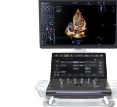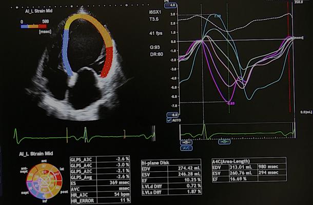
An example of strain imaging cardiac echocardiography software offered on Canon's cardiac ultrasound systems. It is highly automated to speed workflow. It color codes areas of different strain values, graphs these in a visual plot and pulls quantification measurements. Photo by Dave Fornell.
The biggest trend across healthcare the past two years is the emergence of artificial intelligence (AI) and its integration into numerous cardiac ultrasound systems from several vendors. The current AI-enabled echocardiography systems help speed workflow by auto identifying the cardiac anatomy, creating automated quantification, automated ejection fractions and mitral valve regurgitation PISA scores, and the ability to view 3-D datasets and pull out the optimal views for auto measurements to eliminate inter-operator variability.
The next generation will do more with the image data, where currently only a small amount of it is processed to create images. As computer processing speeds continue to increase rapidly, more of the ultrasound data will be able to be processed in real time, and AI will play an increasing role. This may include AI review in the background of the system for radiomic markers for early stage disease or risk factor quantification that are not evident to the human eye. Often, these smart algorithms can help drastically reduce the amount of time needed to make measurements.
“Machine learning is an exciting area that echo is exploring and adapting to,” said Judy Hung, M.D., director of echocardiography, Division of Cardiology, Massachusetts General Hospital. “We are working with a machine learning scientist at MIT on a number of projects, particularly in valve disease. It will certainly help with automation, so it will improve workflow and improve our accuracy and reproducibility in the area of valve disease. The machine learning can help us look at the variability of these exams and tell us what measurements are not important that we can throw out and ignore, and which ones we need to focus on.”
Watch the VIDEO: Artificial Intelligence for Echocardiography at Mass General — Interview with Judy Hung, M.D.
Cardiac Ultrasound Strain Imaging Seeing Increasing Use
Strain imaging, also referred to as speckle or wall motion tracking, has seen a slow-growing increase over the past decade and there were numerous sessions on the topic at ASE. The main focus of cardiac strain use is in serial cardio-oncology echo exams, said strain imaging expert Marielle Scherrer-Crosbie, M.D., Ph.D., director of echocardiography at the Hospital of the University of Pennsylvania, and chair of the 2019 ASE sessions. She said these exams are used to monitor cardiac toxicity levels in cancer patients treated with radiotherapy near the heart (as in breast and lung cancers) or systemic chemotherapy agents. Strain has emerged as the primary way to track myocardial damage so therapy can be modified to preserve cardiac function and prevent heart failure.
“Strain is a very hot topic,” Crosbie said. “It is an index of myocardial function. It seems in many pathologies strain decreases earlier than the ejection fraction and there is a better prognostic valve of the strain than the ejection fraction.”
Watch the VIDEO: An Overview of Echo Strain Imaging — Interview with Marielle Scherrer-Crosbie, M.D.
Point-of-Care Ultrasound Seeing Widespread Expansion
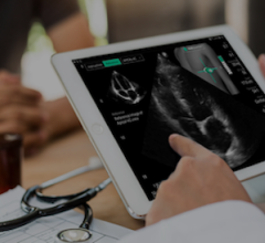
Point-of-care ultrasound (POCUS) has seen a rapid expansion in cardiology offices, emergency rooms and cath labs. POCUS is being used by cardiologists and other physicians for a quick look inside patients as a triage tool to determine if a more detailed echo exam or computed tomography (CT) exam might be needed. There are now several small, pocket or tablet sized POCUS systems. There also are app-based systems that can turn an iPhone into an ultrasound. Many of these systems now integrate automatic left ventricular ejection fraction as a key cardiac diagnostic, so it is consistent and not based on novice user calculations.
Cath labs are seeing rapid adoption of POCUS to image vascular access for better accuracy than anatomical landmarks.
Cath labs also are using ultrasound to aid in procedural navigation, not just with transesophageal echo (TEE), but also as supplemental imaging for peripheral vascular procedures to help reduce amount of X-ray fluoro time.
VIDEO: Point of Care Ultrasound (POCUS) in Cardiology and Critical Care — Interview with Michael Lanspa, M.D.,
Related Echocardiography Trends Content:
Echocardiography Trends at ASE 2019
VIDEO: An Overview of Echo Strain Imaging — Interview with Marielle Scherrer-Crosbie, M.D.
VIDEO: Example of Artificial Intelligence Integrated Into Cardiac Ultrasound
VIDEO: Using an iPhone to Perform a Cardiac Ultrasound Exam
VIDEO: Use of Technology to Address Underserved Populations — Interview with
Partho Sengupta, M.D.
360 Degree View of a Smartphone Performing an Echo Exam
VIDEO: Use of Technology to Address Underserved Populations
VIDEO: Artificial Intelligence for Echocardiography at Mass General — Interview with Judy Hung, M.D.
New Cardiac Imaging Technologies Unveiled at RSNA 2018
5 Key Trends in New Ultrasound Technology
VIDEO: Point of Care Ultrasound (POCUS) in Cardiology and Critical Care — Interview with Michael Lanspa, M.D.,
VIDEO: Editor’s Choice of the Most Innovative Echo Technology at ASE 2018

 April 30, 2026