May 27, 2020 — The Radiological Society of North America (RSNA) announced today that its 106th Scientific Assembly and ...
Advanced Visualization
Software used to manipulate or enhance CT and MRI datasets, including MPR, and 3-D (3D) image reconstruction, perfusion imaging, 3-D printing, and procedural planning and procedural navigation.

 May 27, 2020
May 12, 2020 — Medis acquired Advanced Medical Imaging Development S.r.l. (AMID), a developer and supplier specialized ...
May 12, 2020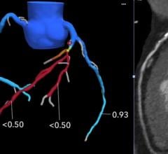
James Udelson, M.D., chief of the division of cardiology, Tufts Medical Center, explains how cardiac computed tomography ...
March 26, 2020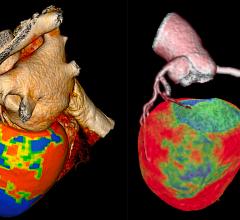
March 16, 2020 — The Society of Cardiovascular Computed Tomography (SCCT) released a new expert consensus document on ...
March 16, 2020
DAIC/ITN Editor Dave Fornell takes a tour of some of the most innovative new medical imaging technologies displayed on ...
January 13, 2020
DAIC Editor Dave Fornell and Imaging Technology News (ITN) Consulting Editor Greg Freiherr offer a post-game report on ...
December 18, 2019
November 27, 2019 — CAE Healthcare will showcase its mixed reality training solutions for practicing physicians and ...
November 27, 2019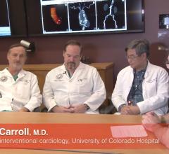
Interview with John Carroll, M.D., director of interventional cardiology, Robert Quaife, M.D., director of advanced ...
October 02, 2019
Feature | Henry Ford Hospital | Dave Fornell, Editor
Henry Ford Hospital thought leaders regularly speak at the cardiology conferences about new research and technology ...
October 01, 2019September 27, 2019 — EchoPixel introduced what it calls the first-ever intraoperative software to provide naked-eye ...
September 27, 2019
September 12, 2019 — HeartFlow Inc. has obtained clearance from the U.S. Food and Drug Administration (FDA) for the ...
September 12, 2019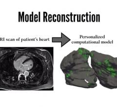
August 19, 2019 — Scientists at Johns Hopkins have successfully created personalized digital replicas of the upper ...
August 19, 2019
An example of Siemens' photo-realistic Cinematic image reconstruction. This image is from a CTA exam from a Siemens ...
August 09, 2019
August 8, 2019 — The Radiological Society of North America (RSNA) and the American College of Radiology (ACR) will ...
August 08, 2019
This is a quick video example of a cardiac computed tomography (CT) exam showing a Medtronic CoreValve transcatheter ...
August 08, 2019