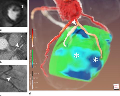
Figure 4 for the study. Images of a 65-year-old man (patient 6). (a) Cardiac MRI perfusion shows perfusion deficit of anterior/anterolateral wall attributed to left anterior descending artery/left circumflex artery (*). (b) CT coronary angiography. (c) Coronary angiography, left anterior oblique projection with caudal angulation. (d) Three-dimensional image fusion helped refine diagnosis: perfusion deficits (*) were most likely caused by narrow first diagonal branch and its first, stented side branch (arrowhead). Retrospectively, denoted lesion could also be found at CT coronary angiography and coronary angiography (arrowheads in b and c, respectively). CT FFR = CT-derived fractional flow reserve, LGE = late gadolinium enhancement. Image courtesy of RSNA, Radiology.
May 4, 2020 – A new technique that combines computed tomography (CT) and magnetic resonance imaging MRI can bolster coronary artery disease diagnosis and help to define appropriate treatment for patients suffering from the disease, according to a new study published in the journal Radiology: Cardiothoracic Imaging.
Coronary artery disease is the most common type of heart disease, according to the Centers for Disease Control and Prevention (CDC). About 18.2 million adults in the United States have coronary artery disease.
CT and MRI are established methods for noninvasive cardiac imaging and evaluation of coronary artery disease. CT is particularly useful for high-resolution images of the coronary anatomy, while cardiac MRI can provide information on blood supply to the heart muscle without exposing patients to ionizing radiation.
Despite their complementary strengths, CT and MRI findings are often analyzed separately, limiting the ability to fully leverage the strengths of the two methods.
“From this experience, the idea came up to fuse information on different pathologic aspects of the disease and to combine them in a single 3D image, which can be interpreted in a very quick but highly accurate fashion,” said study lead author, Jochen von Spiczak, M.D., M.Sc., radiologist and computer scientist at Institute of Diagnostic and Interventional Radiology, University Hospital Zurich in Zurich, Switzerland.
Existing methods of combining CT and MRI have limitations, as they look at only a limited subset of the many aspects of coronary artery disease. Dr. von Spiczak and colleagues overcame these limitations by developing an approach that depicts all the available information from CT and cardiac MRI in one 3D image.
They compared their approach with conventional 2-D readouts in 17 patients who underwent cardiac CT and cardiac MRI due to suspected or known coronary artery disease.
Conventional 2-D readout of the images resulted in uncertain findings in eight cases. The new approach helped solve the divergent findings in six of those cases.
Information from the 3-D fused image helped correlate specific stenoses, or areas of narrowing in the coronary arteries, and their severity with possible cardiac scar tissue and ischemia—a condition in which parts of the heart muscle do not get enough blood. This could be used to help guide interventional or surgical revascularization procedures like stenting or bypass surgery that improve blood supply to the heart.
“The technique may allow for an easier and possibly more accurate identification of patients and coronary stenoses that are likely to benefit from revascularization,” von Spiczak said. “Applying today’s clinical 2-D standard led to a substantial number of uncertain findings in our study, whereas most of these divergent findings could be solved when including additional information from CT-derived blood flow estimates information and 3-D image fusion.”
The study points to a role for the fused approach in complex cases that yield uncertain findings in the first test, such as when results from CT and MRI are inconsistent or even contradictory.
Obstacles to its implementation include higher costs and complexity, problems that may be eased by advances in software, according to von Spiczak.
Reference:

 February 06, 2026