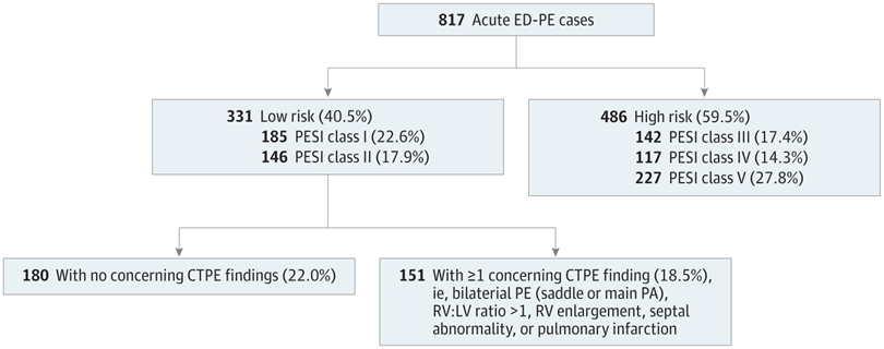
Acute ED-PE cases were divided into low- and high-risk groups based on Pulmonary Embolism Severity Index (PESI) score or class. As in previous reports, cases were spread fairly evenly among the 5 risk classes. A higher proportion was seen in the highest risk class, reflecting the high acuity and rate of comorbidities in the patient population in this study. The low-risk group was further divided based on the presence or absence of 5 concerning findings on pulmonary embolism–protocol computed tomography (CTPE; ie, findings widely believed to confer higher risk). LV indicates left ventricle; PA, pulmonary artery; RV, right ventricle.
June 30, 2023 — Of approximately 250,000 Americans diagnosed with acute pulmonary embolism, or PE, in emergency departments each year, most are hospitalized.
But Michigan Medicine research, published in JAMA Network Open, finds that some patients with PE, a blood clot in one or more pulmonary arteries, may be hospitalized unnecessarily due to computed tomography, or CT, imaging results rather than clinical risk factors.
Approximately 40% of the patients in the study had low risk pulmonary embolism, as defined by the Pulmonary Embolism Severity Index, or PESI score. Roughly half of the low risk patients had CT imaging features that physicians consider “concerning”, and these patients fared just as well in the hospital as those whose CT scans showed no concerning findings.
“The results of this study are going to come as a surprise to most emergency physicians, who have been taught for years to regard large, centrally located, or “saddle”, PEs as true medical emergencies,” said senior author Colin Greineder, M.D., Ph.D., an assistant professor of emergency medicine and pharmacology at University of Michigan Medical School.
“In fact, our findings suggest that CT findings alone might not confer as much risk as we thought. In our population, if the patient’s pulse, rate of breathing, oxygen level and other clinical factors indicated low risk, then ‘concerning’ CT findings by themselves were not associated with worse clinical outcomes.”
Researchers assessed data from more than 800 patients at U-M Health diagnosed with acute pulmonary embolism in the adult emergency department between late 2016 and the end of 2019.
Using the PESI score, patients were divided into low and high risk groups, with the low risk group further divided based on the presence of concerning CT findings. These included large and centrally located PEs, blood clots on both sides of the lung, evidence of strain on the heart due to the blockage and associated infarction, or death, of a region of lung tissue.
Within a month of coming to the emergency department, 18% of patients with high risk PESI scores died, whereas none of the low risk patients with concerning CT imaging findings died. These patients also rarely required intensive care, and their lengths of stay in the hospital were no different than low risk patients without ‘concerning’ CT imaging findings.
“Clinical outcomes were similar, but we found plenty of evidence that emergency physicians are managing these patients differently,” Greineder said. “The rate of discharge and home management was about four times lower in low risk patients with concerning CT imaging findings, and they were more likely to have bedside ultrasounds performed in the emergency department and echocardiograms while in the hospital.”
The study also investigated how often emergency physicians activated the PE Response Team, a multi-disciplinary group designed to make rapid, shared decisions on the management of patients with intermediate and high risk PE at U-M Health.
Response team activation was almost four times more likely in low risk patients with concerning CT imaging findings despite their low risk PESI scores.
Over the past two decades, a strong evidence base has been established to support safe outpatient management of pulmonary embolism, but adoption by emergency medicine clinicians has been slow and remains limited, says co-author Geoffrey Barnes, M.D., M.Sc., associate professor of cardiology-internal medicine at U-M Medical School.
“We have tools like the Pulmonary Embolism Severity Index and biomarkers that allow for appropriate management of these patients, but they do not typically include those ‘concerning’ CT findings. Our report suggests that these imaging findings may be an important barrier to safe discharge.”
The results, researchers say, require further research and confirmation to verify that these findings are replicated in other hospital systems and patient populations.
“Like other groups, we are already working to overcome other barriers to outpatient management of low risk PE,” said first author Connor O’Hare, M.D., an emergency medicine resident at U-M Health. “If our current findings are reproduced in other studies and hospital systems, then we will need effective implementation strategies to overcome the perceived risk associated with ‘concerning’ CT imaging findings in otherwise low risk patients.”
For more information: https://www.uofmhealth.org/

 June 17, 2026


