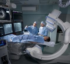
March 8, 2016 ― Fluoroscopy makes guiding a catheter through a blood vessel possible. However, fluoroscopy, a form of real-time moving X-ray, also exposes the patient to radiation. Now, a University of Missouri School of Medicine researcher has evaluated technology that may be used to replace fluoroscopy, eliminating the need for X-ray during cardiac ablation procedures.
“Radiofrequency ablation is a procedure that uses a catheter to administer heat from radio waves to a very small area of the heart to disrupt abnormal signals that cause rhythm problems,” said Sandeep Gautam, M.D., an assistant professor in the Division of Cardiovascular Medicine at the MU School of Medicine and lead author of the study. “Fluoroscopy imaging allows us to pinpoint the treatment area to perform ablation. However, by using fluoroscopy imaging, patients are exposed to radiation ― and the amount varies depending on the procedure. The primary aim of the study was to demonstrate that electroanatomical mapping may be used instead of fluoroscopy to eliminate radiation exposure.”
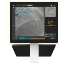
Electroanatomical mapping is a guidance system that creates real-time 3-D maps of the heart without using X-ray technology. For the retrospective study, outcomes of traditional fluoroscopy-guided ablation procedures were compared to the newer mapping system. The two patient groups also were matched for baseline demographics such as age, gender and medical histories.
“We compared the outcomes of an equal number of procedures using both imaging methods,” Gautam said. “For comparison data, we looked at the amount of time it took to place the guidance catheter, the amount of time it took to begin ablation, the duration of ablation and the total length of each procedure.”
Results of the study showed that electroanatomical mapping provided effective imaging equal to fluoroscopy. In addition, there was no statistical difference in any of the comparison times.
“This study builds on previous research to show that there is an alternative to using fluoroscopy during ablation procedures,” Gautam said. “Electroanatomical mapping has evolved to a point that it is comparable to fluoroscopy, yet doesn’t have the risks associated with radiation exposure.”
Although many electrophysiologists continue to use fluoroscopy for ablation procedures due to concerns about procedural duration and efficacy, Gautam feels that change is just around the corner.
“With more research, I think there will be a natural progression toward minimizing and ultimately eliminating the use of fluoroscopy from most cardiac ablation procedures,” Gautam said.
The study, “Fluoroless Radiofrequency Ablation of Typical Cavotricuspid Isthmus-dependent Atrial Flutter is a Safe and Practical Procedure,” recently was published in The Journal of Innovations in Cardiac Rhythm Management.
For more information: www.innovationsincrm.com


 October 24, 2025


