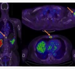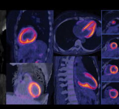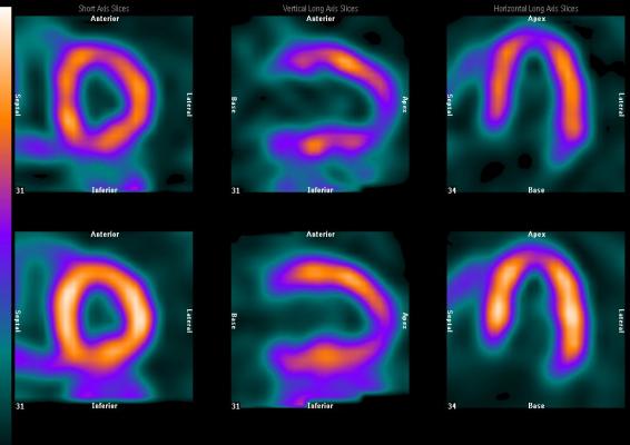
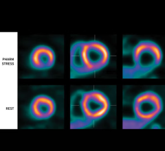
This SPECT/CT study clearly shows ischemia in the heart.
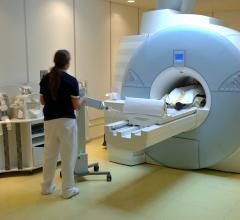
The introduction of hybrid technology — positron emission tomography/computed tomography (PET/CT) and single-photon emission computed tomography (SPECT)/CT -— has revolutionized the imaging world. This technology allows the combination of the exquisite anatomic details provided, for example, by CT, with the important and much needed functional, physiologic or metabolic information provided by molecular imaging.
Hybrid imaging technology has the potential of providing “one-stop” imaging with increased specificity, attenuation correction and localization, thus providing more accurate diagnosis. The utility of PET/CT is established in clinical practice and has added significant value in the areas of neurology, cardiology and oncology. Since the role of PET/CT has been established in clinical practice, the following comments refer to the emerging modality of SPECT/CT.
SPECT/CT is on the Rise
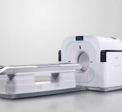
SPECT/CT imaging is gaining more widespread use with increasing clinical applications and availability of equipment. Single-photon emitters are routinely available in nuclear medicine facilities, so imaging is easier to implement, since the equipment is available. Unfortunately, reimbursement for SPECT/CT is lacking at the present time, thus limiting the purchase of equipment and use of the modality.
Guidelines for SPECT/CT performance and interpretation have been established by the Society of Nuclear Medicine and Molecular Imaging.1 The potential applications for SPECT/magnetic resonance imaging (MRI) are exciting and also should enter the clinical practice in near future.
The incremental diagnostic value of SPECT/CT technology, as compared to planar and SPECT imaging alone in a number of clinical situations, including endocrinology, oncology, musculoskeletal, infection imaging and other areas, are impressive and gaining more widespread use.
The following is a look at some specific areas where this technology is being implemented:
Cardiology
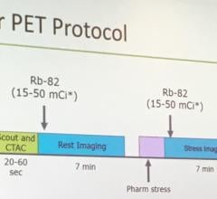
In the area of cardiology, SPECT /CT technology offers attenuation correction at minimum with lower-end systems, thus decreasing breast and diaphragm attenuation artifacts and increasing specificity. The higher-end SPECT/CT systems exemplify the value of “one-stop” imaging technology by combining perfusion, function and coronary anatomy as well as coronary calcium measurement.
Liver/Spleen Imaging
Although most of technetium-99m (Tc-99m) sulfur colloid liver/spleen imaging has been replaced by ultrasound and CT imaging, SPECT/CT offers a significant advantage in evaluation of splenosis, thus excluding more serious diagnosis, such as pancreatic carcinoma, when the accessory splenic tissue cannot be separated by pancreatic tail mass. An article from The European Journal of Nuclear Medicine describes seven of 20 equivocal lesions on ultrasound and MRI correctly identified by SPECT/CT as accessory splenic tissue and confirmed by biopsy.2
Parathyroid Imaging
A number of articles in the literature have demonstrated the incremental diagnostic value of SPECT/CT by accurate localization of parathyroid adenomas in the usual and ectopic location. The study is helpful in decreasing surgery and anesthesia time, thus decreasing overall morbidity. Parathyroid SPECT/CT also offers valuable information for surgical planning (i.e., neck approach versus sternotomy for low and mediastinal adenomas).3
Thyroid Carcinoma
While iodine-131 planar imaging for thyroid cancer imaging is excellent in its ability to diagnose recurrent/metastatic disease, it lacks in accurate localization of disease. Investigators in Europe have shown incremental value of SPECT/CT in 57 percent (41 of 71) cases. Seventeen percent of cases resulted in precise localization of skeletal metastasis, and in six percent pulmonary/mediastinal disease was additionally helpful in equivocal neck uptake.4
Skeletal Imaging: Not Just ‘Hot Spot’ Imaging
The role of SPECT/CT in skeletal evaluation is invaluable. The technology not only offers accurate localization, but also improves specificity with information provided by CT. Benign etiologies such as arthritis trauma can be more accurately interpreted as the etiology of increased activity on bone imaging.
A study reported in the American Journal of Roentenology evaluated a total of 104 osseous lesions. SPECT/CT showed increase in specificity from 19 percent to 81 percent. This would also eliminate the necessity for additional radiographs to further clarify bone scan abnormalities.5-8
Infection Evaluation
In an article published in The Journal of Nuclear Medicine in 2006, the authors studied 82 patients for suspected infection. The incremental value of SPECT/CT was demonstrated in 48 percent of patients. SPECT/CT offered improved diagnostic information, better localization and extent evaluation of infection.9,10
Neuroendocrine Tumors
Indium-111 Octreotide SPECT/CT evaluation in a study consisting of 44 patients showed improved localization in 23, defined extent in 17, detected unsuspected bone lesions in three and was helpful in distinguishing physiologic uptake from pathologic lesion in three patients. In 14 percent of patients, there was an overall change in patient management.11
The incremental diagnostic value has also been shown in other areas, including lymphoscintigraphy and prostascint imaging.
Conclusion
SPECT/CT is rapidly emerging as an important and valuable diagnostic modality offering distinct advantages when compared to planar and SPECT imaging alone. The advantages include better localization, the ability to separate physiologic from pathologic processes and detect unsuspected disease, and the potential for a change in management in some patients. It also offers guidance for surgical approach and decreases surgical and anesthesia time, resulting in both decreased morbidity and costs.
The strength of CT attenuation offered by SPECT/CT, especially in cardiac studies, improves specificity and accuracy. This emerging modality offers increased diagnostic accuracy by combining anatomy and function in one sitting, thus offering state-of–the-art and accurate diagnosis. itn
Lalitha Ramanna, M.D., FACNM, serves as director of nuclear medicine at Providence Little Company of Mary Hospital in Torrance, Calif., and is clinical associate professor at the Keck School of Medicine at the University of Southern California, Los Angeles. Ramanna is the president of the Society of Nuclear Medicine and Molecular Imaging’s (SNMMI) Correlative Imaging Council and sits on the board of directors for SNMMI’s PET Center of Excellence.
References
1. Delbeke D, Coleman RE, Guiberteau MJ, et al. “Procedure guideline for SPECT/CT imaging1.0.” J Nucl Med. 2006; 47:1227-1234.
2. Horger M, Eschmann SM, Lengerke C, Claussen CD, Pfannenberg C, Bares R. “Improved detection of splenosis in patients with haematological disorders: the role of combined transmission-emission tomography.” Eur J Nucl Med Mol Imaging. 2003; 30(2):316-9. Epub 2002 Dec 14.
3. Lavely WC, Goetze S, Friedman KP, et al. “Comparison of SPECT/CT, SPECT, and planar imaging with single- and dual-phase 99mTc-sestamibi parathyroid scintigraphy.” J Nucl Med. 2007; 48:1084–1089.
4. Tharp K, Israel O, Hausmann J, et al. “Impact of 131I-SPECT/CT images obtained with an integrated system in the follow-up of patients with thyroid carcinoma.” Eur J Nucl Med Mol Imaging. 2004; 31:1435–1442.
5. Even-Sapir E, Flusser G, Lerman H, Lievshitz G, Metser U. “SPECT/multislice low-dose CT: a clinically relevant constituent in the imaging algorithm of nononcologic patients referred for bone scintigraphy.” J Nucl Med. 2007; 48:319–324.
6. Roemer W, Noemayr A, Uder M, Bautz W, Kuwert T. “SPECT-Guided CT for evaluating foci of increased bone metabolism classified as indeterminate on SPECT in cancer patients.” J Nucl Med. 2006; 47:1102-1106.
7. Utsunomiya D, Shiraishi S, Imuta M, et al. “Added value of SPECT/CT fusion in assessing suspected bone metastasis: comparison with scintigraphy alone and nonfused scintigraphy and CT.” Radiology. 2006; 238:264–271.
8. Horger M, Eschmann SM, Pfannenberg C, et al. “Evaluation of combined transmission and emission tomography for classification of skeletal lesions.” AJR. 2004; 183:655–661.
9. Filippi L, Schillaci O. “SPECT/CT with a hybrid camera: a new imaging modality for the functional anatomical mapping of infections.” Expert Rev Med Devices. 2006; 3:699-703.
10. Bar-Shalom R, Yefremov N, Guralnik L, et al. “SPECT/CT using 67Ga and 111In-labeled leukocyte scintigraphy for diagnosis of infection.” J Nucl Med. 2006; 47:587–594.
11. Krausz Y, Keidar Z, Kogan I, et al. “SPECT/CT hybrid imaging with 111In-pentetreotide in assessment of neuroendocrine tumours.” Clin Endocrinol (Oxf). 2003; 59:565-73.
 June 23, 2025