
Pamela Woodard, M.D., led a team that designed a new imaging agent that may light up dangerous plaque in arteries.
January 30, 2015 — The U.S. Food and Drug Administration (FDA) has approved for human evaluation a nanoparticle-based imaging agent jointly developed at Washington University School of Medicine in St. Louis and the University of California, Santa Barbara, in collaboration with Texas A&M University. The imaging agent may illuminate dangerous plaque in arteries, and doctors hope to use it to identify patients at high risk of stroke.
“This is the first receptor-targeted nanoparticle agent for cardiovascular imaging approved for investigational use in humans,” said principal investigator Pamela K. Woodard, M.D., professor of radiology and of biomedical engineering. “Starting with bench research, then developing and testing the agent and taking it through the FDA process into human patients has involved an extensive team of basic scientists, clinical researchers and clinicians.”
In patients with atherosclerosis, plaque accumulates on the inner walls of arteries that deliver blood to the body.
“Plaque is a complex structure made up of cholesterol, calcified deposits and other substances, all of which can cause inflammation,” said Woodard, also director of the Center for Clinical Imaging Research at the Mallinckrodt Institute of Radiology at Washington University. “Depending on the severity of the inflammation, these plaques can be stable or progress to a vulnerable phase in which they rupture, leading to stroke or heart attack.”
According to Woodard, many studies have indicated that most patients with plaque narrowing a carotid artery won’t go on to have a stroke.
“With current technology — such as ultrasound — we can’t tell whether the plaque is vulnerable or stable,” she said. “So we can’t distinguish the high-risk patients who need surgery from low-risk patients who can be treated with medication alone. We designed this nanoparticle agent to develop a test that can detect these vulnerable plaques and identify those patients at highest risk of stroke and in need of surgery to remove the plaque.”
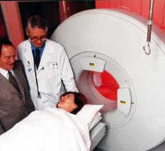
This nanoparticle agent illuminates plaque in any of the body’s arteries and can be detected with a positron emission tomography (PET) scan. Researchers recently began testing the safety of the nanoparticle in healthy individuals. They next will focus on patients with atherosclerosis who already are scheduled to undergo surgery to remove plaque from their carotid arteries.
“In this way, we’ll be able to see whether the areas that light up in the image because of our nanoparticles are the same areas that contain vulnerable plaque, as assessed from the surgeries,” Woodard said. “Once we show success imaging the carotid arteries, we will evaluate the nanoparticle agent in other vessels such as the coronary arteries, which represent a greater challenge because of their smaller size and complex motion.”
The nanoparticle also carries copper atoms, making it visible with a standard PET scanner. Similar small amounts of copper-64 regularly are used in PET scans, a technique common in cancer detection and therapy and neurologic imaging.
In addition, components of the nanoparticle also are designed to self-assemble in the watery environment of blood.
“The success of this nanoparticle system relies on the controlled self-assembly of functional polymers in water, which is driven by the careful design of hydrophilic (water-attracting) and hydrophobic (water-repelling) segments into the polymers,” said Craig J. Hawker, Ph.D., professor and director of the California Nanosystems Institute at the University of California, Santa Barbara.
Added Woodard: “We have been able to develop this highly receptor-specific imaging technology because of the generous support from the National Heart, Lung and Blood Institute, our diverse and dedicated team of investigators and our extensive facilities that allow us to make this nanoparticle imaging agent in a sterile environment, meeting the FDA requirements for use in people.”
According to the university’s Office of Technology Management, Pamela K. Woodard, Geoffrey E. Woodard, Rafaella Rossin and the late Michael J. Welch are the inventors of the patented NPR-C imaging method.
For more information: www.medicine.wustl.edu


 November 17, 2025


![Phase III clinical trial of [18F]flurpiridaz PET diagnostic radiopharmaceutical meets co-primary endpoints for detecting Coronary Artery Disease (CAD)](/sites/default/files/styles/content_feed_medium/public/Screen%20Shot%202022-09-13%20at%203.30.13%20PM.png?itok=2w6OoNd6)