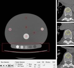
July 15, 2020 — Cardiac CT exams performed to assess heart health also provide an effective way to screen for osteoporos ...
Computed Tomography (CT)
Cardiac computed tomography CT systems use a series of X-ray images to create an image volume dataset that can be sliced or manipulated on any plane using advanced visualization software. This channel includes content on CT scanners, CT contrast agents, CT angiography (CTA and CCTA), CT perfusion, spectral CT (also called dual souce or dual energy CT), and interative image reconstruction software that can reduce dose and make lower-quality CT images diagnostic.

Feature | FFR Technologies | Dave Fornell, Editor
Fractional flow reserve (FFR) pressure wires have been used now in interventional cardiology procedures for more than a ...
 July 07, 2020
Canon Medical Systems USA has created a virtual trade-show experience for its cardiovascular computed tomography (CT) ...
July 06, 2020Sponsored Content

Feature | Cardiac Imaging
Sponsored Content — According to the American Heart Association, cardiovascular disease is the leading cause of death in ...
March 13, 2026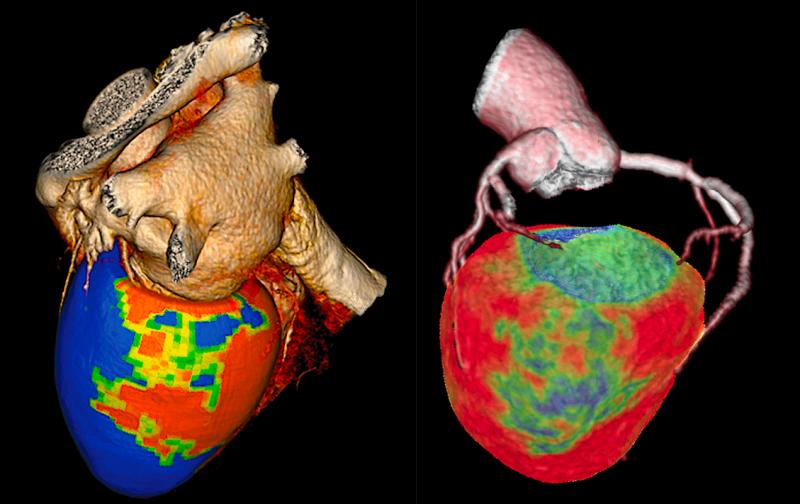
Feature | CT Angiography (CTA) | Dave Fornell, Editor
Here is an overview of a few of the biggest technology advances in cardiovascular computed tomography (CT). These are ...
June 08, 2020
May 27, 2020 — The Radiological Society of North America (RSNA) announced today that its 106th Scientific Assembly and ...
May 27, 2020May 12, 2020 — Medis acquired Advanced Medical Imaging Development S.r.l. (AMID), a developer and supplier specialized ...
May 12, 2020Sponsored Content

Blog | Enterprise Imaging
As medical advancements continue to push the boundaries of what is possible in the field of structural heart ...
August 10, 2023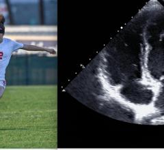
May 11, 2020 – Competitive athletes are a rapidly growing population worldwide. Habitual vigorous exercise, a defining ...
May 11, 2020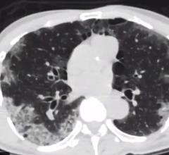
Interview with Geoffrey Rose, M.D., president of Sanger Heart and Vascular Institute with Atrium Health, in Charlotte ...
May 07, 2020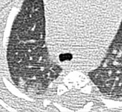
May 5, 2020 — Children and teenagers with COVID-19 showed distinctive clinical and computed tomography (CT) findings ...
May 05, 2020
May 5, 2020 — The Society of Cardiovascular Computed Tomography (SCCT) announced plans this week to hold its annual ...
May 05, 2020
May 4, 2020 – A new technique that combines computed tomography (CT) and magnetic resonance imaging MRI can bolster ...
May 04, 2020
April 23, 2020 — A special report published in the journal Radiology outlines prevention, diagnosis and treatment of ...
April 30, 2020
Stephen Bloom, M.D., FASNC, director of nonivasive cardiology (cardiac CT, nuclear cardiology and echocardiography) at M ...
April 18, 2020
Feature | Coronavirus (COVID-19) | Dave Fornell, Editor
Cases of acute cardiovascular disease and cardiac complications caused by COVID-19 require cardiovascular imaging ...
April 18, 2020
Hicham Skali, M.D., a staff cardiologist and member of the Non-invasive Cardiovascular Imaging Program at Brigham and ...
April 04, 2020