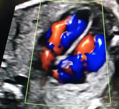
Dia's artificial intelligence automation or point-of-care cardiac ultrasound on display at the 2021 Radiological Society of North America (RSNA) annual meeting.
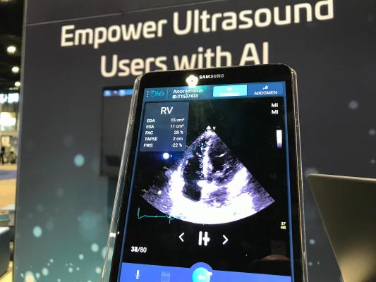
December 2, 2021 — Artificial intelligence (AI) vendor DiA Imaging Analysis was featured in a recent study presented by a team of cardiac physicians from Mount Sinai’s Icahn School of Medicine at the American Heart Association (AHA) 2021 virtual meeting. The breakthrough study demonstrated the accuracy of LVivo RV, Dia's AI for right ventricle function analysis, validating its sensitivity and specificity results compared to Cardiac MRI (CMR).
Right Ventricle (RV) size and function are vital indicators of cardiac dysfunction, including pulmonary embolism (PE), pulmonary hypertension, and right heart failure. Amid the COVID-19 pandemic, cardiac ultrasound imaging includes assessment of the RV to detect RV dysfunction, commonly seen in patients with COVID-19. Although common, RV function analysis using an ultrasound device is challenging and often requires additional examination due to the RV’s challenging geometry.
The recent study of 125 patients compared DiA’s right heart AI-based software to CMR. Results demonstrated high sensitivity and specificity when compared to CMR, the gold standard of cardiac imaging. The study concluded that DiA’s automated AI-based right heart analysis for automated RV function quantification has the potential to accurately and instantly detect evidence of right heart dysfunction at the echo lab and near the bedside.
“As echo is the first imaging triage modality used for scanning suspected heart issues, this study validated the potential of adding AI as part of our cardiac ultrasound workflow,” said Martin Goldman, M.D., professor of cardiology at Mount Sinai and one of the authors of the study.
“We are proud to share that leading physicians from the Icahn School of Medicine at Mount Sinai, a globally ranked program, have validated our FDA-approved AI software,” said Hila Goldman-Aslan, CEO and co-founder of DiA Imaging Analysis. “The study demonstrated that LVivo RV can provide an automated and accurate solution to the critical analysis of the right ventricle function using ultrasound.”
The study, Validation of Artificial Intelligence for Assessing Right Ventricular Function by Transthoracic Echocardiography,[1] was conducted by Brian Hsia, Ashton Lai, Rajeev Samtani, Solomon Bienstock, Steve Liao, Eric Stern, Gina LaRocca, Javier Sanz, Stamatios Lerakis, Gregg W Stone, Lori Croft, and Martin Goldman, of the Icahn School of Medicine at Mount Sinai, New York, N.Y. The results were presented to the American Heart Association on November 15, 2021. The study was published in Circulation Magazine, Volume 144, Issue 1.
DiA also demonstrated its suite of AI-based ultrasound software solutions at RSNA 2021.
DiA Imaging Analysis has FDA cleared and CE marked ultrasound AI software solutions that make the use and analysis of ultrasound images smarter, faster, and accessible to all. The company's LVivo product line for cardiac and abdominal automated analysis allows clinicians with various levels of ultrasound experience to use and analyze ultrasound images on their ultrasound devices or healthcare IT systems with increased speed, efficiency and accuracy. DiA serves thousands of end-users worldwide.
For more information: www.dia-analysis.com
Reference:

 October 08, 2025