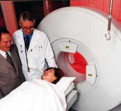
December 28, 2009 - The Medical College of Wisconsin will license a novel molecular imaging technology designed to rapidly diagnosis cell death in organs such as the brain and heart.
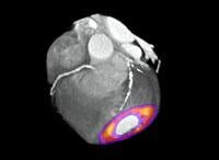
The technology, using imaging probes with a radiopharmaceutical compound, was invented by Ming Zhao, Ph.D., assistant professor of biophysics. The probes bind to dead and dying cells making them useful for detecting acute cell injury and cell death. When the active component of this molecule is attached to a radioactive tracer, it can be used in nuclear medicine imaging techniques, such as positron emission tomography (PET) or single photon emission computed tomography (SPECT), to produce three-dimensional images of where this cell death is occurring.
"Imaging agent discovery and development is an important aspect in molecular and medical imaging research," Zhao said. "The process is critical for the improvement of existing imaging technologies and for early detection of acute cell death, cancerous tissue growth and major vessel diseases."
He said the ability to image dead and dying cells can have major clinical benefits. For example, it could allow oncologists to rapidly monitor tumor response to a specific therapy. Another potential application is for rapid diagnosis of myocardial infarction. Often patients come into ER complaining of chest pain and need to have an expensive overnight hospital stay so they can be monitored while their lab results are being processed. This compound could allow clinicians to noninvasively image the heart and determine within a few hours if the patient actually had a heart attack or something else.
The university announced today it signed a licensing agreement with GE Healthcare to further evaluate and develop the technology with the option to commercialize it.
For more information: www.mcw.edu

 November 17, 2025


![Phase III clinical trial of [18F]flurpiridaz PET diagnostic radiopharmaceutical meets co-primary endpoints for detecting Coronary Artery Disease (CAD)](/sites/default/files/styles/content_feed_medium/public/Screen%20Shot%202022-09-13%20at%203.30.13%20PM.png?itok=2w6OoNd6)