September 14, 2020 — Philips Healthcare released new imaging and workflow enhancements for its novel Kodex-EPD cardiac imaging and mapping system. The system is now being used to treat patients with atrial fibrillation (AFib) at 40 sites worldwide with over 1,500 patients treated.
The latest release includes improvements in electrophysiology (EP) imaging and mapping performance, as well as enhanced features to support cryoablation procedures.
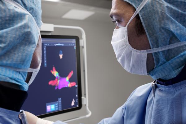
Unlike traditional methods to image the heart during AF procedures such as CT and X-ray fluoroscopy, the Kodex-EPD system uses a combination of sensors attached to the body and catheters inside the heart to image the anatomy and properties of the heart, using innovative dielectric sensing technology. As a result, electrophysiologists can create detailed 3-D views of the heart of a patient in as little as three minutes and navigate to the target location more easily and efficiently, with the required precision and without using radiation.
“The new release of the Kodex-EPD system represents a significant step forward in terms of image quality and workflow efficiency for AF procedures, as we continue to work towards our longer term goal of providing real-time therapy assessment,” said Marlou Janssen, general manager of Philips EPD solutions. “By partnering with Medtronic and developing unique capabilities for cryoablation, we can give more physicians access to this truly innovative cardiac imaging and mapping system, and contribute to delivering efficient workflows with Medtronic’s best-in-class cryo-ablation therapy.”
Advances in the new release include faster, high-resolution imaging, improvements in mapping functionality and point density, as well as new visualization options such as ‘multi-chamber’ view to help understand the relative positions of adjacent chambers and ‘glass view’, which provides physicians with an improved perception of 3-D catheter location and orientation within the heart. For cryoablation procedures, the Kodex-EPD system offers enhanced Occlusion assessment functionality with high accuracy and a simplified workflow.
“Using the Kodex-EPD imaging system’s ability to provide high resolution imaging, revealing specific pulmonary vein morphologies, has allowed me to personalize my ablation approach in Medtronic cryoballoon procedures,” said Marcin Kowalski M.D., director of electrophysiology at Staten Island University Hospital, N.Y. “Using the two innovative technologies together has significant benefits for our patients.”
“Combining the short procedure times of cryoablation with the real-time imaging capabilities of the Kodex-EPD system, we have been able to further improve our efficient lab workflow” said Professor Lukas Dekker, electrophysiologist at Catharina Hospital in Eindhoven, the Netherlands. “Moreover, fluoroless imaging with the Kodex-EPD system may reduce the dependency on fluoroscopy and pre-procedural CTs and thus help to reduce X-ray exposure to patients and staff, which is important to us.”
The Kodex-EPD cardiac imaging and mapping system is commercially available in the U.S., Europe, and China. It is part of Philips’ unique portfolio of systems, smart devices, software and services in image-guided therapy, which combine to provide healthcare providers with sophisticated, procedure-oriented solutions.
For more information: www.philips.com/epd
Related Kodex-EDP EP Mapping System Content:
Northwestern Medicine One of First to Use Dielectric Imaging System to Guide Cardiac Ablation
4 Key Takeaways From the 2019 Heart Rhythm Society Meeting
Philips and Medtronic Collaborate on Image-guided Atrial Fibrillation Treatment

 May 22, 2026


