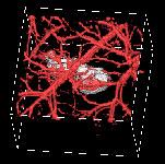
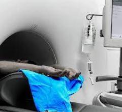
A example of a photoacoustic image.
May 4, 2009 - The Society of Nuclear Medicine (SNM) Symposium on Multimodality Cardiovascular Molecular Imaging convened April 30 and May 1 at the National Institutes of Health (NIH) in Bethesda, Md., to discuss the prevention of cardiac events as a critical next step toward improving cardiovascular health and patient care.
The meeting was hosted by SNM’s Molecular Imaging Center of Excellence (MICoE) with the support of NIH and of multiple corporate and nonprofit partners.
“The symposium was designed to help disseminate the latest cardiovascular imaging research and raise awareness of the most promising clinical techniques with the potential to decrease the frequency and mitigate the severity of heart attacks and other cardiac events,” said MICoE President Henry F. VanBrocklin, Ph.D., professor of radiology and director of radiopharmaceutical research at the University of California, San Francisco.
Speakers from the fields of chemistry, engineering, physics, molecular biology, cardiovascular physiology and imaging sciences focused on important lessons that can be gleaned from imaging damaged heart tissue in hopes of diagnosing potential cardiac events and preventing them before they occur.
“Changes in the structure, geometry and eventually function of the left ventricle occur following myocardial infarction, or heart attack,” said Albert J. Sinusas, M.D., professor of medicine and diagnostic radiology and director of cardiovascular nuclear imaging, Yale School of Medicine. “A targeted molecular imaging approach can provide both prognostic and diagnostic potential by revealing unique insights into molecular processes involved in repair and remodeling of the heart following a heart attack.”
During the symposium, speakers presented research about new imaging and drug delivery technologies that are currently being developed to counter the effects of atherosclerosis, or chronic inflammation of the heart’s arterial walls. According to these researchers, cardiovascular disease is the leading cause of mortality and morbidity in developed countries and atherosclerosis is a major cause of severe cardiovascular disease.
“One third of adult men and women have some form of cardiovascular disease and the estimated direct and indirect costs to our health care system exceed $400 billion,” said David K. Glover, Ph.D., associate professor of cardiovascular medicine, University of Virginia School of Medicine. “We are currently investigating an antibody-based molecular imaging probe that may be a useful and sensitive tool for advancing our understanding of the formation of atherosclerotic lesions and for the noninvasive detection of vulnerable plaques.”
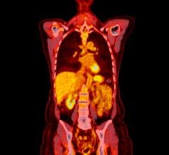
Other presentations explored both the established and the leading edge in imaging techniques, from positron emission tomography (PET) and magnetic resonance (MR), to a variety of hybrid imaging modalities in biomedicine, including applications of ultrasound for medicine and nondestructive testing.
“Metabolic imaging has played a key role in diagnosis and prognostication as well as directing therapy and understanding these disease states and new therapies,” said Robert S. B. Beanlands, M.D., director of the University of Ottawa Heart Institute’s cardiac PET center. “A recent comprehensive meta-analysis confirmed that FDG PET is the most sensitive method available for predicting heart wall motion recovery and revealing improved outcomes in the healthy heart wall tissue of patients whose blood supply has been restored.”
“Photoacoustic imaging represents one of the most promising techniques for molecular imaging,” said Matthew O’Donnell, Ph.D., the Frank and Julie Jungers dean of engineering, professor of bioengineering and adjunct professor of mechanical engineering, College of Engineering at the University of Washington. “Photoacoustics combines optical and acoustic methods and can provide real-time images with molecular sensitivity at significant image depth with high spatial resolution.”
For more information: www.snm.org

 November 17, 2025


![Phase III clinical trial of [18F]flurpiridaz PET diagnostic radiopharmaceutical meets co-primary endpoints for detecting Coronary Artery Disease (CAD)](/sites/default/files/styles/content_feed_medium/public/Screen%20Shot%202022-09-13%20at%203.30.13%20PM.png?itok=2w6OoNd6)