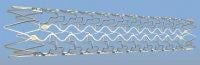
June 1, 2009 - Six months after placement of a vProtect Luminal Shield to treat a "vulnerable plaque,” a follow-up exam revealed reduced plaque burden and reduced stenosis in the target vessel.
The exam was part of the SECRITT I trial, a randomized study designed to evaluate the vProtec Luminal Shield as a prophylactic treatment for nonflow-limiting vulnerable plaques. The trial is sponsored and conducted by Erasmus University in Rotterdam. The six-month follow-up exam was performed May 20 by an interventional cardiology team led by Dr. Patrick Serruys, the principal investigator for the SECRITT I study. Evelyn Regar, M.D., Ph.D., moderated the case at the EuroPCR congress in Barcelona.
The vProtect Luminal Shield is manufactured by Prescient Medical, which plans to pursue a vulnerable plaque indication in the future.
The patient, a 75-year-old woman, was treated six months earlier for a culprit lesion in the left anterior descending coronary artery. At that time the SECRITT investigators, using a combination of imaging techniques, also located a vulnerable plaque in the patient's left circumflex artery. The lesion was unremarkable by angiography (37 percent stenosis); such lesions are usually not treated, even though vulnerable plaques are prone to rupture with potentially fatal consequences. A 3.5 mm vProtect Luminal Shield was placed in the vessel (reference vessel diameter 2.74 mm), resulting in a post-procedural stenosis of 30 percent.
The six-month angiographic follow-up exam was performed per study protocol using a variety of imaging techniques: multislice CT, intravascular ultrasound (IVUS), IVUS-virtual histology (IVUS-VH), angiography, and optical coherence tomography (OCT). Additional details on the SECRITT I study methodology can be found in a case study published recently in Nature Reviews Cardiology (Volume 6, May 2009).
The follow-up exam indicated the self-expanding vProtect Luminal Shield had stabilized the vulnerable lesion while gently expanding over time. Imaging studies revealed that the residual stenosis in the area of the lesion improved from 30 percent to just 5 percent (measured by quantitative angiography). The shield was covered by a thin layer of healthy tissue with an average thickness of 80-110 microns.
“These results are comparable to what is seen with drug-eluting stents in terms of thickness. We did not see a significant hyperplastic injury response,” said Gary Tearney, M.D., Ph.D., cardiac pathologist, associate professor of pathology at Harvard University and the Wellman Center for Photomedicine. “The Shield appears to be perfectly designed for just this type of lesion-since it’s self-expanding, there is less chance of rupturing the plaque.”
For more information: www.prescientmedical.com
 April 25, 2023