April 27, 2017 — The vacancy rate for radiographers increased to 4.2 percent in 2017, according to the latest American ...
Cardiac Imaging
The cardiac imaging channel includes the modalities of computed tomography (CT), cardiac ultrasound (echocardiography), magnetic resonance imaging (MRI), nuclear imaging (PET and SPECT), and angiography.

 April 27, 2017
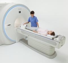
April 24, 2017 — Offering a full range of advanced clinical applications for researchers, Toshiba Medical will ...
April 24, 2017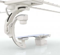
April 18, 2017 — Patients at Terrebonne General Medical Center (TGMC) in Houma, La., now have access to safe, high ...
April 18, 2017Sponsored Content

Feature | Cardiac Imaging
Sponsored Content — According to the American Heart Association, cardiovascular disease is the leading cause of death in ...
March 13, 2026
Feature | Cardiac Imaging | Dave Fornell
As part of U.S. healthcare reform efforts, starting Jan. 1, 2018, physicians will be required to document they are ...
April 18, 2017
Feature | CT Angiography (CTA) | Dave Fornell
Cardiac computed tomography (CT) imaging really took off a decade ago with the introduction of 64-slice scanners, which ...
April 13, 2017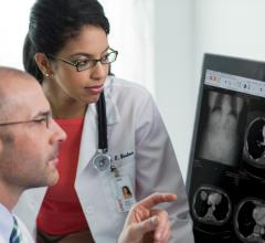
April 5, 2017 — The Society of Cardiovascular Computed Tomography (SCCT) and Toshiba Medical announced a new partnership ...
April 05, 2017Sponsored Content

Feature
SPONSORED CONTENT — Studycast is a comprehensive imaging workflow system that allows healthcare professionals to work ...
June 17, 2025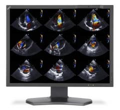
April 3, 2017 — The American Society of Echocardiography (ASE) and the European Association of Cardiovascular Imaging ...
April 03, 2017
April 3, 2017 — The newest release of American College of Radiology (ACR) Appropriateness Criteria covers 230 topics ...
April 03, 2017
DAIC Editor Dave Fornell takes a tour of some of the interesting new technologies from the vendor booths on the expo ...
April 03, 2017Sponsored Content

Feature | Cardiac Imaging
Cardiac positron emission tomography (PET) is growing in popularity among cardiologists because it provides the ability ...
March 05, 2024
Feature | Dave Fornell
April 3, 2017 — Here is the list of the top 25 most popular pieces of content on the Diagnostic and Interventional ...
April 03, 2017
MDBuyline analyst Tom Watson shares some of the most important trends in cardiac technology he saw at the 2017 American ...
March 28, 2017
March 24, 2017 — At the 2017 American College of Cardiology's Annual Scientific Session & Expo (ACC.17), Philips ...
March 24, 2017Sponsored Content

Blog | Enterprise Imaging
As medical advancements continue to push the boundaries of what is possible in the field of structural heart ...
August 10, 2023
March 21, 2017 — Toshiba Medical demonstrated its Forward projected model-based Iterative Reconstruction SoluTion (FIRST ...
March 21, 2017
March 16, 2017 — During the 66th Annual Scientific Session & Expo of the American College of Cardiology (ACC), March 17 ...
March 16, 2017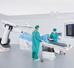
March 15, 2017 — The U.S. Food and Drug Administration (FDA) has cleared the Artis pheno robotic C-arm angiography syste ...
March 15, 2017