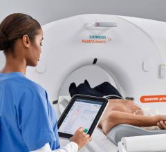
March 14, 2019 — Siemens Healthineers will introduce the Somatom go.Top Cardiovascular Edition, a new version of its ...
Computed Tomography (CT)
Cardiac computed tomography CT systems use a series of X-ray images to create an image volume dataset that can be sliced or manipulated on any plane using advanced visualization software. This channel includes content on CT scanners, CT contrast agents, CT angiography (CTA and CCTA), CT perfusion, spectral CT (also called dual souce or dual energy CT), and interative image reconstruction software that can reduce dose and make lower-quality CT images diagnostic.
 March 14, 2019
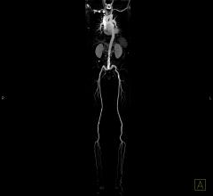
March 13, 2019 — An imaging procedure commonly performed before starting cancer treatment can provide valuable clues ...
March 13, 2019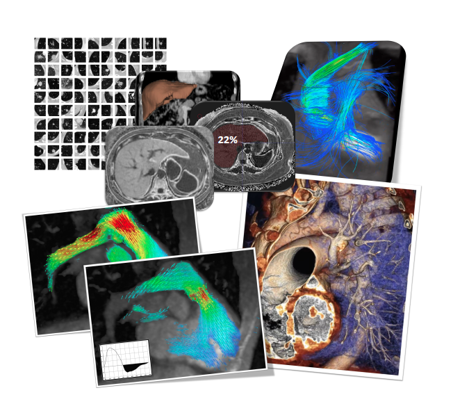
Feature | Cardiac Imaging | By Greg Freiherr
Computed tomography (CT) and magnetic resonance imaging (MRI) scans are chock full of information that might be used to ...
March 12, 2019Sponsored Content

Feature | Cardiac Imaging
Sponsored Content — According to the American Heart Association, cardiovascular disease is the leading cause of death in ...
March 13, 2026
Can computed tomography (CT) angiography accurately measure fractional flow reserve (FFR)? Even if it can, is CT-derived ...
March 08, 2019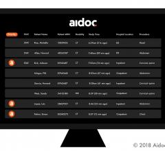
March 4, 2019 — Artificial intelligence (AI) radiology solution provider Aidoc announced the commercial release of its ...
March 04, 2019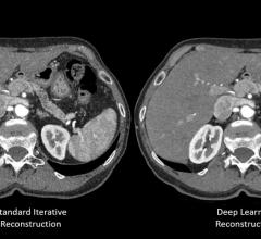
February 27, 2019 — Canon Medical Systems recently introduced AiCE (Advanced intelligent Clear IQ Engine), a deep ...
February 27, 2019Sponsored Content

Blog | Enterprise Imaging
As medical advancements continue to push the boundaries of what is possible in the field of structural heart ...
August 10, 2023
February 13, 2019 — At the 2019 Healthcare Information and Management Systems Society (HIMSS) global conference and ...
February 13, 2019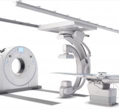
February 6, 2019 — Canon Medical Systems USA Inc. recently introduced a new angiography configuration featuring its ...
February 06, 2019
DAIC Editor Dave Fornell takes a tour of some of the most interesting new medical imaging technologies displayed on the ...
January 26, 2019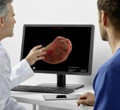
January 25, 2019 — Siemens Healthineers presented its first intelligent software assistant for radiology, the AI-Rad ...
January 25, 2019
January 8, 2019 — The Society of Cardiovascular Computed Tomography (SCCT) has released a new expert consensus document ...
January 08, 2019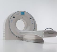
December 12, 2018 — In stroke, time saved on imaging is time gained in the treatment window. The recently updated ...
December 12, 2018
November 30, 2018 – Clinical diagnostics intelligence platform company MaxQ AI and Samsung NeuroLogica announced a ...
November 30, 2018
November 28, 2018 — Vital, a Canon Group company, will highlight the latest additions to its enterprise imaging portfoli ...
November 28, 2018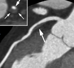
November 27, 2018 – Zebra Medical Vision and Clalit Health Services announced the completion of a research project that ...
November 27, 2018