June 20, 2019 — Bay Labs announced that new data on the company’s first-of-its-kind deep learning investigational ...
Ultrasound Imaging
Ultrasound uses sound waves to create images. This section includes echocardiography (echo), transthoracic echo (TTE), transesophageal echo (TEE), echo contrast, transducers, ultrasound software and point of care (POC) ultrasound.
 June 20, 2019
June 19, 2019 — DiA Imaging Analysis announced the presentation of two studies assessing the performance and accuracy of ...
June 19, 2019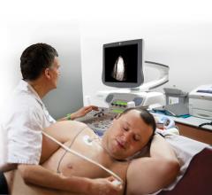
As part of the Consolidated Appropriations Act of 2018, pass-through payment status for LUMASON® (sulfur hexafluoride ...
May 29, 2019Sponsored Content
Case Study | Cardiovascular Ultrasound
As part of the Consolidated Appropriations Act of 2018, pass-through payment status for LUMASON® (sulfur hexafluoride ...
May 29, 2019
Physicians use many strategies to better interface with patients and their families to try and explain in non-physician ...
May 28, 2019
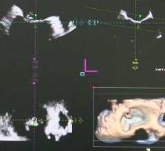
This is an example of how the heart's left atrial appendage (LAA) can be evaluated for thrombus and possible ...
May 16, 2019
This is an example of a carotid artery reporting module from Change Healthcare at 2018 Radiological Society of North ...
May 16, 2019
Feature | Artificial Intelligence | Ross Upton
Artificial Intelligence has a multitude of impacts on our daily lives, from recommending movies based upon your Netflix ...
May 07, 2019
Alex Haak, Ph.D., clinical scientist at Philips Health Systems North America, is based at the University of Colorado ...
May 01, 2019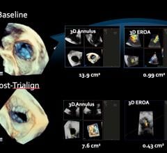
April 30, 2019 — A new document compiled by four cardiac imaging professional societies provides a resource to guide ...
April 30, 2019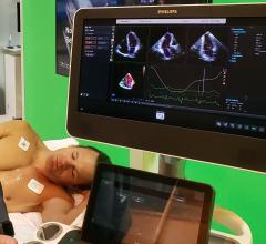
April 26, 2019 — With healthcare costs continuing to rise, affordable and accurate imaging and diagnosis achieved ...
April 26, 2019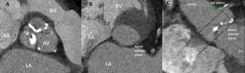
Feature | Structural Heart | Nadeen N. Faza, M.D., Dee Dee Wang, M.D., Joao Cavalcante, M.D., Andrew D. Choi, M.D., Jeffrey B. Geske, M.D., and Stephen H. Little, M.D.
Recent months have signaled a new and exciting era in the dynamic world of structural heart disease (SHD). The COAPT ...
April 24, 2019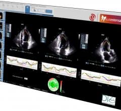
April 16, 2019 — DiA Imaging Analysis has partnered with the Italian healthcare IT company Ebit (Esaote Group), to offer ...
April 16, 2019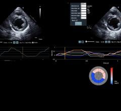
April 9, 2019 — DiA Imaging Analysis announced the launch of LVivo SAX, a cardiac analysis tool that helps clinicians ...
April 09, 2019
Feature | Cardiovascular Ultrasound | Dave Fornell, Editor
Echocardiography reporting systems are usually integrated with, or offered as an add-on module for a cardiovascular ...
April 08, 2019© Copyright Wainscot Media. All Rights Reserved.
Subscribe Now