American Society of Nuclear Cardiology (ASNC) President Dennis Calnon, M.D., MASNC, FASE, FSCCT, director of cardiac ...
PET Imaging
Positron emission tomography (PET) is a nuclear imaging technology (also referred to as molecular imaging) that enables visualization of metabolic processes in the body. The basics of PET imaging is that the technique detects pairs of gamma rays emitted indirectly by a positron-emitting radionuclide (also called radiopharmaceuticals, radionuclides or radiotracer). The tracer is injected into a vein on a biologically active molecule, usually a sugar that is used for cellular energy. PET systems have sensitive detector panels to capture gamma ray emissions from inside the body and use software to plot to triangulate the source of the emissions, creating 3-D computed tomography images of the tracer concentrations within the body.

 February 01, 2022
American Society of Nuclear Cardiology (ASNC) President Dennis Calnon, M.D., MASNC, FASE, FSCCT, director of cardiac ...
February 01, 2022
October 6, 2021 – A new study published in Radiology: Cardiothoracic Imaging on cardiac imaging trends over a decade ...
October 06, 2021Sponsored Content

Feature | Cardiac Imaging
Cardiac positron emission tomography (PET) is growing in popularity among cardiologists because it provides the ability ...
March 05, 2024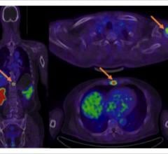
July 13, 2021 — In a recent blog, the American Society of Nuclear Cardiology (ASNC) reported that Humana, one of the ...
July 13, 2021
Feature | Nuclear Imaging | By Staff of the American Society of Nuclear Cardiology (ASNC)
A year after COVID-19 turned the world upside down, the American Society of Nuclear Cardiology (ASNC) asked members how ...
June 02, 2021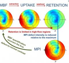
April 1, 2021 – The ability to measure myocardial blood flow (MBF) as part of myocardial perfusion imaging (MPI) is one ...
April 01, 2021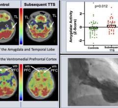
March 31, 2021 — Heightened activity in the brain, caused by stressful events, is linked to the risk of developing a ...
March 31, 2021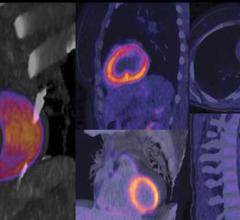
Ernest Garcia, Ph.D., MASNC, FAHA, endowed professor in cardiac imaging, director of nuclear cardiology R&D laboratory ...
September 25, 2020
Feature | Coronavirus (COVID-19) | Dave Fornell, Editor
Cases of acute cardiovascular disease and cardiac complications caused by COVID-19 require cardiovascular imaging ...
April 18, 2020
Hicham Skali, M.D., a staff cardiologist and member of the Non-invasive Cardiovascular Imaging Program at Brigham and ...
April 04, 2020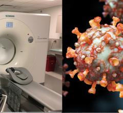
News | Coronavirus (COVID-19) | Dave Fornell, Editor
April 3, 2020 — A new guidance document on best practices to maintain safety and minimize contamination in nuclear ...
April 03, 2020
As hospital imaging departments look to replace aging nuclear scanners with updated technology, many are asking if ...
February 19, 2020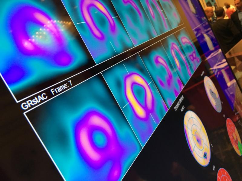
Feature | Nuclear Imaging | Dave Fornell, Editor
There were a few key takeaways from the American Society of Nuclear Cardiology (ASNC) 2019 annual meeting in September ...
November 22, 2019 imaging program. She spoke on this topic at the 2019 meeting of the American Society Nuclear Cardiology (ASNC) and led a tour with attendees of the PET-CT system at Rush, which was located close to the conference. #ASNC")
This is a photo essay of new technologies and activities at the American Society of Nuclear Cardiology (ASNC) 2019 ...
November 11, 2019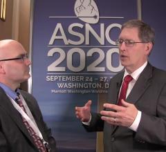
Rob Beanlands, M.D., FASNC, 2019 American Society of Nuclear Cardiology (ASNC) president, shares a couple trends he sees ...
November 07, 2019