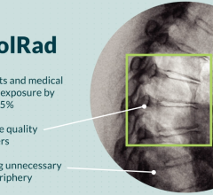
November 5, 2021 – An FDA-approved device used during cardiac cath lab procedures cut radiation exposure for ...
Cardiac Imaging
The cardiac imaging channel includes the modalities of computed tomography (CT), cardiac ultrasound (echocardiography), magnetic resonance imaging (MRI), nuclear imaging (PET and SPECT), and angiography.
 November 05, 2021
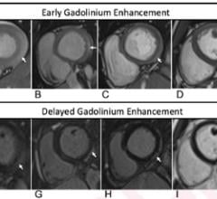
November 1, 2021 — According to ARRS’ American Journal of Roentgenology (AJR), radiologists need to be cognizant of the ...
November 01, 2021
Feature | By Dave Fornell, DAIC Editor
November 1, 2021 — Here is the list of the most popular content on the Diagnostic and Interventional Cardiology (DAIC) ...
November 01, 2021Sponsored Content

Feature | Cardiac Imaging
Sponsored Content — According to the American Heart Association, cardiovascular disease is the leading cause of death in ...
March 13, 2026
Feature | Cardiac Diagnostics | By Dave Fornell, DAIC Editor
October 29, 2021 — A new guideline for the evaluation and diagnosis of chest pain was released this week that provides ...
October 29, 2021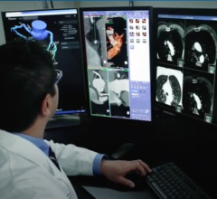
October 20, 2021 — HeartFlow Inc. announced the commencement of the REVEALPLAQUE (A pRospEctiVe, multicEnter study to ...
October 20, 2021
October 14, 2021 — Cardiac computed tomography angiography (CTA) derived left atrium emptying fraction (LAEF) improves ...
October 14, 2021Sponsored Content

Feature
SPONSORED CONTENT — Studycast is a comprehensive imaging workflow system that allows healthcare professionals to work ...
June 17, 2025
October 6, 2021 — Data presented during the late-breaking science session at the European Society of Cardiology (ESC) 20 ...
October 06, 2021
October 6, 2021 – A new study published in Radiology: Cardiothoracic Imaging on cardiac imaging trends over a decade ...
October 06, 2021
October 4, 2021 – UltraSight, a digital health company developing artificial intelligence (AI) enabled cardiac imaging ...
October 04, 2021Sponsored Content

Feature | Cardiac Imaging
Cardiac positron emission tomography (PET) is growing in popularity among cardiologists because it provides the ability ...
March 05, 2024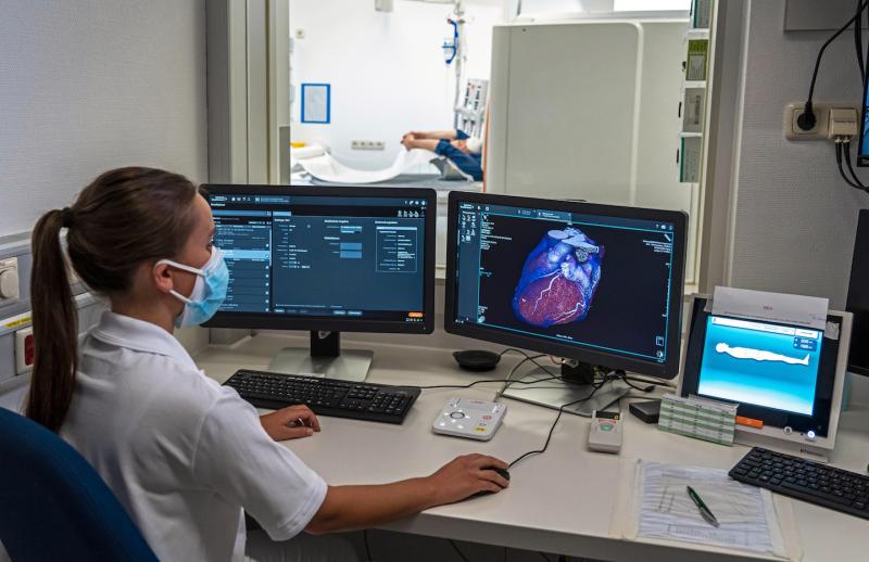
Feature | Computed Tomography (CT) | By Dave Fornell, DAIC Editor
The U.S. Food and Drug Administration (FDA) Sept. 30 cleared the world's first photon-counting computed tomography (CT) ...
October 04, 2021![Test selection should be a shared decision between patient and physician rather than directed by insurers’ test substitution policies, according to a statement published online in the Journal of the American College of Cardiology.[1] The statement summarizes the proceedings of a recent summit convened by the American Society of Nuclear Cardiology (ASNC), leadership of the American College of Cardiology Imaging Council, American Society of Echocardiography (ASE), Society of Cardiovascular Computed Tomography](/sites/default/files/styles/content_feed_medium/public/Cardiac_imaging_Nuclear_echo_CT_MRI.jpg?itok=XH9oijOU)
September 22, 2021 — Test selection should be a shared decision between patient and physician rather than directed by ...
September 22, 2021
September 14, 2021 – Us2.ai, a Singapore-based medtech firm backed by Sequoia India and EDBI, has received U.S. Food and ...
September 14, 2021Sponsored Content

Blog | Enterprise Imaging
As medical advancements continue to push the boundaries of what is possible in the field of structural heart ...
August 10, 2023
September 13, 2021 — Early coronary angiography in out-of-hospital cardiac arrest (OHCA) patients without ST-segment ...
September 13, 2021
Feature | Artificial Intelligence | By Dave Fornell, Editor
Artificial intelligence (AI) is growing in all areas of medicine and was the topic of several advanced technology ...
September 08, 2021
One of the trends in cardiovascular information system (CVIS) and radiology PACS at the Healthcare Information ...
August 31, 2021