October 20, 2021 — HeartFlow Inc. announced the commencement of the REVEALPLAQUE (A pRospEctiVe, multicEnter study to ...
Computed Tomography (CT)
Cardiac computed tomography CT systems use a series of X-ray images to create an image volume dataset that can be sliced or manipulated on any plane using advanced visualization software. This channel includes content on CT scanners, CT contrast agents, CT angiography (CTA and CCTA), CT perfusion, spectral CT (also called dual souce or dual energy CT), and interative image reconstruction software that can reduce dose and make lower-quality CT images diagnostic.
 October 20, 2021
October 14, 2021 — Cardiac computed tomography angiography (CTA) derived left atrium emptying fraction (LAEF) improves ...
October 14, 2021
October 6, 2021 — Data presented during the late-breaking science session at the European Society of Cardiology (ESC) 20 ...
October 06, 2021Sponsored Content

Feature | Cardiac Imaging
Sponsored Content — According to the American Heart Association, cardiovascular disease is the leading cause of death in ...
March 13, 2026
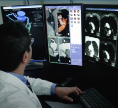
October 6, 2021 – A new study published in Radiology: Cardiothoracic Imaging on cardiac imaging trends over a decade ...
October 06, 2021
Feature | Computed Tomography (CT) | By Dave Fornell, DAIC Editor
The U.S. Food and Drug Administration (FDA) Sept. 30 cleared the world's first photon-counting computed tomography (CT) ...
October 04, 2021
September 27, 2021 — Zebra Medical Vision, the deep-learning medical imaging analytics company, announces its eighth U.S ...
September 27, 2021Sponsored Content

Blog | Enterprise Imaging
As medical advancements continue to push the boundaries of what is possible in the field of structural heart ...
August 10, 2023![Test selection should be a shared decision between patient and physician rather than directed by insurers’ test substitution policies, according to a statement published online in the Journal of the American College of Cardiology.[1] The statement summarizes the proceedings of a recent summit convened by the American Society of Nuclear Cardiology (ASNC), leadership of the American College of Cardiology Imaging Council, American Society of Echocardiography (ASE), Society of Cardiovascular Computed Tomography](/sites/default/files/styles/content_feed_medium/public/Cardiac_imaging_Nuclear_echo_CT_MRI.jpg?itok=XH9oijOU)
September 22, 2021 — Test selection should be a shared decision between patient and physician rather than directed by ...
September 22, 2021
Feature | Cardiovascular Ultrasound | By Dave Fornell, Editor
Outside of medicine, computer-generated virtual twins of real machines like cars or airplanes have been used in ...
July 26, 2021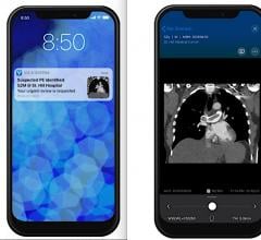
July 21, 2021 — Artificial intelligence (AI) medical imaging vendors Viz.AI and Avicenna.AI have partnered to enable ...
July 21, 2021
July 16, 2021 — Canon Medical Systems USA is joining forces with Cleerly in a strategic partnership to support simple ...
July 16, 2021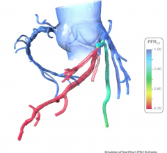
July 15, 2021 — HeartFlow, which has commercialized noninvasive computed tomography derived fractional flow reserve (FFR ...
July 15, 2021
July 14, 2021 — Performing the first cardiac scan on their new photon-counting detector computed tomography (CT) scanner ...
July 14, 2021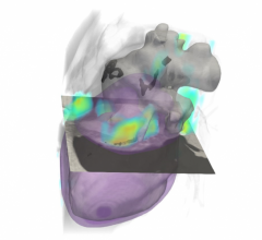
June 14, 2021 — Heart disease and cancer are the leading causes of death in the United States, and it’s increasingly ...
June 14, 2021
June 3, 2021 — Medical imaging AI specialist Avicenna.AI announced that it has received certification in the United ...
June 07, 2021
May 27, 2021 — Philips Healthcare released a workhorse computed tomography (CT) system, the Spectral CT 7500, which has ...
May 27, 2021