
The researchers developed an ultra thin monolithic OCT endoscope that overcomes the limitations of current, larger OCT catheters by using 3-D printed components.
October 1, 2020 — A team of researchers led by the University of Adelaide and University of Stuttgart has used 3-D micro-printing to develop the world’s smallest, flexible optical coherence tomography (OCT) imaging catheter for looking inside blood vessels. A multidisciplinary team of researchers and clinicians was able to 3-D print a tiny lens on to the end of an optical fibre, the thickness of a human hair.
There work was published in the journal Light: Science & Applications.[1]
The camera-like imaging device can be inserted into blood vessels to provide high quality 3-D images to help scientists better understand the causes of heart attack and heart disease progression, and could lead to improved treatment and prevention. The imaging device is so small that researchers were able to scan inside the blood vessels of mice.
The researchers developed an ultrathin monolithic OCT endoscope that overcomes the limitations of current, larger OCT catheters by using two-photon polymerization to 3-D print 125 μm diameter micro-optics directly onto the optical fiber inside the the catheter. Freeform micro-optics have been created for correcting the nonchromatic aberrations of highly miniaturized probes, which cannot be fabricated using traditional techniques. The ultrathin OCT endoscope achieved a measured full width at half maximum (FWHM) focal spot size of 12.4 μm and effective depths of focus (the depth range in which FWHM < 2FWHMmin29) of 760 µm (x axis) and 1,100 µm (y axis). The utility of the ultrathin endoscope is demonstrated on both in situ preclinical (mouse) and ex vivo clinical (human) models of cardiovascular disease. We are now able to reveal details of the tissue microarchitecture at depths not previously achieved with such small imaging probes. To the best of our knowledge, this is the smallest aberration-corrected intravascular probe to have been developed, the researchers explained.
Dr. Jiawen Li, co-author and Heart Foundation Post-doctoral Fellow at the Institute for Photonics and Advanced Sensing, University of Adelaide, said in Australia cardiovascular disease kills one person every 19 minutes.
“A major factor in heart disease is the plaques, made up of fats, cholesterol and other substances that build up in the vessel walls,” Li said. “Preclinical and clinical diagnostics increasingly rely on visualizing the structure of the blood vessels to better understand the disease. Miniaturized endoscopes, which act like tiny cameras, allow doctors to see how these plaques form and explore new ways to treat them,” she said.
Dr. Simon Thiele, group leader, optical design and simulation at the University of Stuttgart, was responsible for fabricating the tiny lens.
“Until now, we couldn’t make high quality endoscopes this small,” Thiele said. “Using 3D micro-printing, we are able to print complicated lenses that are too small to see with the naked eye. The entire endoscope, with a protective plastic casing, is less than half a millimeter across."
“It’s exciting to work on a project where we take these innovations and build them into something so useful," Li explains. “It’s amazing what we can do when we put engineers and medical clinicians together."
The research collaboration also included researchers from The South Australian Health and Medical Research Institute, The Royal Adelaide Hospital and Monash University.
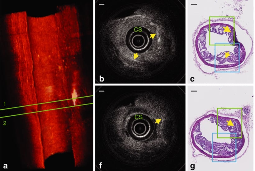
A: Three-dimensional rendering of the volumetric data set acquired with a 3-D printed intravascular imaging catheter in a diseased mouse artery. The volume comprises 258 frames of OCT images. 2. This rendering reveals distributions of cholesterol crystals, which are indicated by white. B: Cross-sectional OCT image of region 1 in A. C: corresponding histology image. F: Cross-sectional OCT image of region 2 in A. G: corresponding histology image; Scale bar equals 100 µm. Find more images and information.
Related 3-D printing Content:
The Future of 3-D Printing in Medicine
World’s Smallest Intravascular OCT Imaging Device Created Using 3-D Printing
Researchers 3-D Print a Beating Heart From Human Cells
New Technique Allows More Complicated 3-D Bioprinting
The Use of 3-D Printing in Cardiology
Reference:

 June 18, 2026


