May 19, 2020 — Atrial disease has been implicated in embolic stroke of undetermined source (ESUS) and a late-breaking ...
Magnetic Resonance Imaging (MRI)
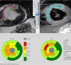
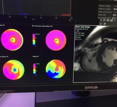
Cardiac MRI creates images from the resonance of hydrogen atoms when they are polarized to face in one direction and then hit with an electromagnetic pulse to knock them off axis. The wobbling of the atoms is what is recorded by computers and used to reconstruct the images. Cardiac MR allows very detailed visualization of the myocardial tissue above the resolution found with cardiac CT. Using different protocol sequences, various contrast type images can be created with MRI to enhance various tissues or to provide physiological data on the function of the heart. This section includes MR analysis software, MRI scanners, gadolinium contrast agents, and related magnetic resonance accessories.
May 12, 2020 — Medis acquired Advanced Medical Imaging Development S.r.l. (AMID), a developer and supplier specialized ...
May 11, 2020 – Competitive athletes are a rapidly growing population worldwide. Habitual vigorous exercise, a defining ...
As medical advancements continue to push the boundaries of what is possible in the field of structural heart ...
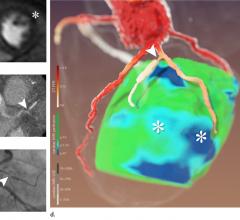
May 4, 2020 – A new technique that combines computed tomography (CT) and magnetic resonance imaging MRI can bolster ...
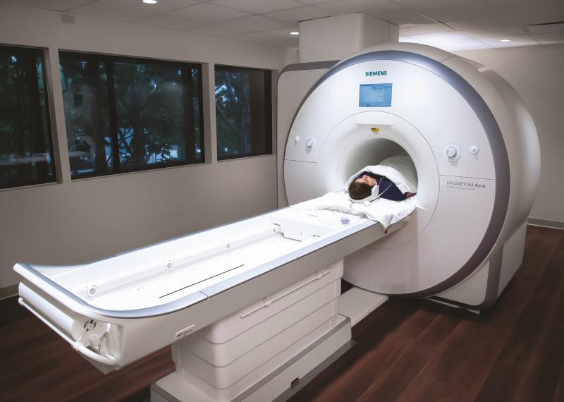
Magnetic resonance imaging (MRI) has been described for the past few decades as a futuristic imaging technology that ...
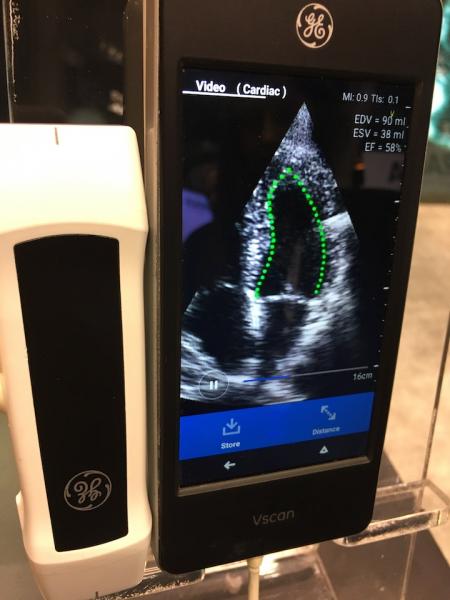
February 7, 2020 – At the 2019 Radiological Society of North America (RSNA) meeting in December, there was a record ...
Karen Ordovas, M.D., MAS, professor of radiology and cardiology at the University of California San Francisco (UCFS) ...
James Carr, M.D., chair of the Department of Radiology, Northwestern University, and incoming 2020 President of the Soci ...
October 28, 2019 — Leaders within University Hospitals and the Harrington Heart and Vascular Institute had a vision to ...
October 21, 2019 — Elevated left ventricular mass, known as left-ventricular hypertrophy, is a stronger predictor of ...
October 16, 2019 — Researchers have shown for the first time in preclinical studies that the drug Aliskiren can delay ...
October 7, 2019 – A recent study published in the New England Journal of Medicine supports the use of cardiac magnetic ...
September 25, 2019 – Cardiac magnetic resonance imaging (MRI) analysis can be performed significantly faster with ...
September 9, 2019 — The American Society of Nuclear Cardiology (ASNC) published a new expert consensus document along ...

Traditionally, computed tomography (CT) and ultrasound have been the workhorse imaging modalities in the world of ...

 May 19, 2020
