Rami Doukky, M.D., system chair, Division of Cardiology, professor of medicine, Cook County Health and Hospitals System ...
Nuclear Imaging
Nuclear imaging, also called molecular imaging, includes positron emission computed tomography (PET) and single photon emission computed tomography (SPECT) imaging. This section includes radiopharmaceuticals and tracers, PET-CT, SPECT-CT, and PET-MRI.

 June 29, 2017
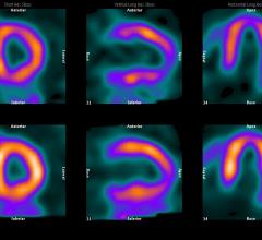
June 15, 2017 — The American Society of Nuclear Cardiology (ASNC) has published an updated consensus statement on ...
June 15, 2017Feature | Radiation Dose Management | Dominic Siewko
In recent years, radiation dose management awareness has heightened across the healthcare industry to address growing ...
June 13, 2017Sponsored Content

Feature | Nuclear Imaging
Cardiac PET/CT represents a major advancement in cardiovascular diagnostics, offering significant clinical and ...
November 12, 2025
May 31, 2017 — MIM Software Inc. recently announced significant updates to its MIM Encore solution for viewing nuclear ...
May 31, 2017May 25, 2017 — At the 2017 annual meeting of the Society for Imaging Informatics in Medicine (SIIM), June 1-3 in ...
May 25, 2017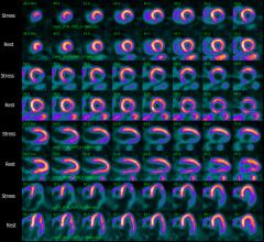
May 22, 2017 — Lantheus Holdings Inc., parent company of Lantheus Medical Imaging Inc., and GE Healthcare announced the ...
May 22, 2017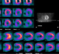
Study Reveals Low Adoption of IAEA Recommendations for Reduced Nuclear Cardiology Radiation Exposure
May 12, 2017 — A study in 65 countries has revealed low adoption of International Atomic Energy Agency (IAEA) ...
May 12, 2017
May 11, 2017 — A large nuclear cardiology laboratory in Missouri has slashed its average radiation dose by 60 percent in ...
May 11, 2017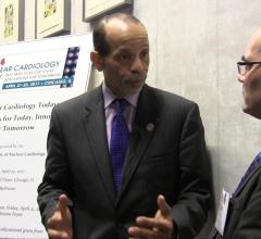
Kim A. Williams, Sr., M.D., chief of cardiology at Rush University Medical Center, Chicago and former president of both ...
May 03, 2017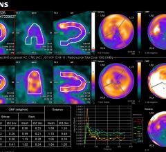
April 28, 2017 — Researchers at the Australian Nuclear Science and Technology Organisation (ANSTO) have led the ...
April 28, 2017
David Wolinsky, M.D., director of nuclear cardiology at Cleveland Clinic Florida and past-president of the American ...
April 28, 2017
Feature | Cardiac Imaging | Dave Fornell
As part of U.S. healthcare reform efforts, starting Jan. 1, 2018, physicians will be required to document they are ...
April 18, 2017
April 13, 2017 — The University of Missouri Research Reactor (MURR) and its partners Nordion and General Atomics (GA) ...
April 13, 2017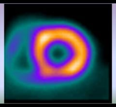
April 11, 2017 — IBA Molecular announced that it has merged with previous acquisition Mallinckrodt Nuclear Medicine LLC ...
April 11, 2017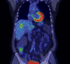
April 5, 2017 — GE Healthcare has signed an agreement with HealthTrust, a group purchasing organization headquartered in ...
April 05, 2017