Videos

Cardiac Imaging

Cardiovascular Ultrasound | July 20, 2022

Enhanced features on the Philips EPIQ CVx ultrasound system provide a next level photorealistic 3D rendering, making it easy for users to interpret what they are seeing. The TrueVue feature enhances the sense of depth and space, producing images that appear natural and realistic to the human eye.  

Watch the video to check out how TrueVue can help improve views of LAA and MV morphology. 

Cardiovascular Ultrasound | July 07, 2022

Automated features on the Philips EPIQ CVx cardiology ultrasound system are helping to bring consistency and speed to every echo exam. The AI-empowered algorithm delivers fast and consistent measurements – in half the time of manual methods.*  

Watch the video to see how you can put “smart”  to work with the latest AI-powered quantification tools. 

*External study with external sonographers comparing the results of 18 exams with and without AutoMeasure

Cardiovascular Ultrasound | June 14, 2022

The X5-1c transducer from Philips provides enhanced clinical information in transthoracic imaging over a standard phased array transducer. When combined with nSIGHT Plus image formation on the EPIQ CVx cardiology ultrasound system, the X5-1c transducer enables image quality rarely seen from a transthoracic transducer. 

Watch the video to learn about the benefits which may include decreased exam time due to faster access to echo windows, increased confidence in quantification results and more. 

Cardiovascular Ultrasound | May 31, 2022

Philips recently announced an update to their flagship EPIQ CVx premium cardiology ultrasound system. Watch the video to learn about the latest features including the next-generation nSIGHT Plus imaging architecture system that leverages both hardware and software to support sophisticated image formation and enhanced image quality.

Cardiac Imaging | February 01, 2022

Interview with Campbell Rogers, M.D., chief medical officer of HeartFlow which has developed a CT image-based fractional flow reserve (FFR-CT) algorithm. The technology was recently included as a recommendation for front line chest pain evaluations in the 2021 ACC/AHA chest pain evaluation guidelines.

The new guidelines gave high levels of evidence for the use of computed tomography and FFR-CT cardiac imaging as front line imaging modalities for chest pain evaluation.

Related Chest Pain Imaging Content:

First International Chest Pain Diagnosis Guidelines Released

VIDEO: Why the ASNC Did Not Endorse the 2021 Chest Pain Guidelines — Interview with ASNC President Dennis Calnon, M.D.


Coronavirus (COVID-19) | February 01, 2022

Campbell Rogers, M.D., chief medical officer of HeartFlow, explains how hospitals are using CT image-based fractional flow reserve (FFR-CT) assessments to speed throughput of patients in emergency rooms and reduce the need for diagnostic angiograms during the COVID-19 pandemic.


Cardiac Imaging | February 01, 2022

American Society of Nuclear Cardiology (ASNC) President Dennis Calnon, M.D., MASNC, FASE, FSCCT, director of cardiac imaging for McConnell Heart Hospital at Riverside Methodist Hospital, and director of nuclear imaging for Ohio Health Heart and Vascular Physicians in Columbus, explains why the society did not endorse the first-ever U.S. or international guideline for the evaluation and diagnosis of patients with acute or stable chest pain.

Nuclear myocardial perfusion imaging (MPI) has been a gold-standard for evaluation of coronary disease in patients for years, and it is included in the new guidelines. However, computed tomography angiography (CTA) has seen immense growth over the past decade and gained a prominent position in the guidelines as a front line imaging modality. This is due to nearly all hospital emergency rooms now having access to CT systems capable of performing immediate cardiac exams. CT has been seen mainly as an anatomical imaging test, but it also can be used for myocardial perfusion imaging using iodine contrast. While CT has had limits with its ability to image through heavily calcified vessels or stents, that is changing with new CT technologies now coming into service.

One new CT technology that is prominently included I the new chest pain guidelines is CT fractional flow reserve (FFR-CT) imaging.  This uses a computational fluid dynamics algorithm to analyze a patient's CTA. It sends back a report and an interactive 3D reconstruction of all the coronary vessels that shows a color coded drop in FFR ratios, which is a measure of blood flow. Past a certain threshold, the reduced flow needs to be treated with revascularization. Lower level blockages can be treated with drug therapies. FFR-CT is widely expected in the coming years to become a gate keeper for invasive diagnostic angiograms. The hope is it will eliminate the need for the majority of cath lab angiogram exams and only send patients to the lab that need a stent or angioplasty. 

However, the ASNC had major issues with FFR-CT being included in the chest pain guidelines. Its board members argued there is need for more evidence and there should have been more information contraindications for its use, which was included on other imaging modalities in the guidelines. They also argued access to the technology has been very limited. Calnon explains more detail in the video on why this was a sticking point and caused the ASNC to not endorse the guidelines.

Read more in the article — First International Chest Pain Diagnosis Guidelines Released

Nuclear Imaging | February 01, 2022

American Society of Nuclear Cardiology (ASNC) President Dennis Calnon, M.D., MASNC, FASE, FSCCT, director of cardiac imaging for McConnell Heart Hospital at Riverside Methodist Hospital, and director of nuclear imaging for Ohio Health Heart and Vascular Physicians in Columbus, offers a concise overview of new technologies that are enhancing cardiac nuclear imaging. He also explains some of the new developments and uses for PET and SPECT imaging technology.

Find more nuclear imaging technology news


Coronavirus (COVID-19) | January 25, 2022

Siddharth Singh, M.D., director of the COVID-19 heart program, staff cardiologist and echocardiographer, Cedars-Sinai Hospital, explains what has been learned in the first year of the hospital's long-COVID clinic. Cedar-Sinai was one of the first hospitals in the U.S. to create a cardiac long-COVID clinic. As of January 2022, the clinic has been more than 120 long-COVID patients with cardiac specific complains. 

"Depending on what study you read, a percentage of patients with acute COVID infection will go on to develop long-lasting sequelae, but typically the percentage falls over time," explained Siddharth Singh, M.D., director of the COVID heart program, staff cardiologist and echocardiographer, Cedars-Sinai Hospital.

He said studies show between 10-25 percent of COVID patients will experience at least one disabling long-COVID symptom six months after their acute infection.

Cedars-Sinai long-COVID has seen over 500 patients since late 2020. Of these, 120 were referred for cardiac evaluations. The most common presentations in those patients include:
   • Shortness of breath
   • Exertional intolerance
   • Chest pain
   • Heart palpitations  
   • Sensations of light-headedness and dizziness
   • Insomnia
   • Brain fog 
   • Constipation
   • Diarrhea
   • Numbness or tingling in the extremities
   • Longer lasting issues with smell and taste

Singh also said many patients experience anxiety and depression due to the continued symptoms, and are concerned the issues they have from COVID may be permanent. Both Baggish and Trivax also reported seeing these issues in many of their patients.

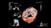
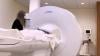
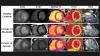
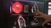
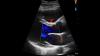
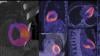
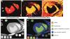
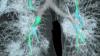
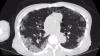
In a smaller subset of patients these clinics have found lingering pericarditis or myocarditis. Most of these patients reported developing chest pain within two week or so after their acute infection. Singh said abnormalities can be imaged using echo and MRI.

"They do typically have abnormalities on imaging. But reassuringly, of all the patients who came to our clinic with these issues, we did not see any decline in left ventricular ejection fraction or systolic function," Singh stressed.

Patients with lingering pericarditis or myocarditis issues are treated with anti-inflammatories. Singh reinforced that the number of patients treated for this have been very low.

Singh said the arrhythmias he has seen include atrial fibrillation (AF) and supraventricular tachycardia. He noted ventricular tachycardia is very rare in these patients. In discussions with his electrophysiology (EP) colleagues, Singh believes the issues with post-COVID arrhythmias often comes down to the substrate of the patient's heart. He said the more cardiovascular disease, cardiomyopathy, scarring, or previous AF a patient has, the more predisposed they are to developing an arrhythmia after a COVID infection. 

Watch more of the interview with Singh in the VIDEO: Examination of Cardiac COVID Long-Haulers
 

Related Long-COVID Content:

What We Know About Cardiac Long-COVID Two Years Into the Pandemic 

VIDEO: Long-term Cardiac Impacts of COVID-19 Two Years Into The Pandemic — Interview with Aaron Baggish, M.D.

VIDEO: Long-COVID Presentations in Cardiology at Beaumont Hospital — Interview with Justin Trivax, M.D.

VIDEO: Examination of Cardiac COVID Long-Haulers — Interview with Siddharth Singh, M.D.

Find more COVID news and video


Coronavirus (COVID-19) | January 25, 2022

Siddharth Singh, M.D., director of the COVID-19 heart program, staff cardiologist and echocardiographer, Cedars-Sinai Hospital, explains how they work up cardiac patients in the Cedars-Sinai long-COVID clinic. He goes through the list of exams  and labs they use in these patients.

Cardiac patients at the Cedar-Sinai long-COVID clinic are evaluated with a basic physical exam, extensive family and medical history, an ECG, basic labs that include troponin and BNP, D-dimer, A1C, lipid panel and a metabolic profile since many patients have become much less active during the pandemic. Patients with symptoms are evaluated with an echocardiogram, including global longitudinal strain. If patients have abnormalities on the echo, or if there is suspected pericarditis or myocarditis, the patient is referred for a cardiac MRI with contrast. 

Watch more of this interview in the VIDEO: Cardiac Presentations in COVID Long-haulers at Cedars-Sinai Hospital 

Related Long-COVID Content:

What We Know About Cardiac Long-COVID Two Years Into the Pandemic 

VIDEO: Long-term Cardiac Impacts of COVID-19 Two Years Into The Pandemic — Interview with Aaron Baggish, M.D.

VIDEO: Long-COVID Presentations in Cardiology at Beaumont Hospital — Interview with Justin Trivax, M.D.

VIDEO: Cardiac Presentations in COVID Long-haulers at Cedars-Sinai Hospital — Interview with Siddharth Singh, M.D.

Find more COVID news and video


Magnetic Resonance Imaging (MRI) | January 18, 2022

Orlando Simonetti, Ph.D., professor, cardiovascular medicine, worked with Siemens to help develop a new, lower-field magnetic resonance imaging (MRI) system, the Magnetom Free.Max. It can scan patients that previously may have been contraindicated because of implantable medical devices. The first system installed in the U.S. is at The Ohio State University Wexner Medical Center. It has a much lower magnetic field and a larger patient opening, removing barriers to MRI imaging for many patients.

Simonetti and his colleagues developed new techniques to boost the signal-to-noise ratio in MRI machines, which allowed the creation of a machine with a lower magnetic field strength that still enables high quality images.

The system gained FDA clearance in July 2021 and was featured by Siemens at the Radiological Society of North America (RSNA) 2021 meeting.

The interview and footage was provided by The Ohio University State University Wexner Medical Center.

Read more in the article New FDA-approved MRI Expands Access to Life-saving Imaging.


Artificial Intelligence | January 13, 2022

Here are two examples of artificial intelligence (AI) driven pulmonary embolism (PE) response team apps featured by vendors Aidoc and Viz.AI at the 2021 Radiological Society of North America (RSNA) 2021 meeting.

The AI scans computed tomography (CT) image datasets as they came off the imaging system and looked for evidence of PE. If detected by the algorithm, it immediately sends an alert to the stroke care team members via smartphone messaging. This is done before the images are even loaded into the PACS. The radiologist on the team can use a link on the app to open the CT dataset and has basic tools for scrolling, windowing and leveling to determine if there is a PE and the severity. The team can then use the app to send messages, access patient information, imaging and reports. This enabled them all to be on the same page and can communicate quickly via mobile devices, rather than being required to use dedicated workstations in the hospital. 

Both vendors showed similar apps for stroke at RSNA 2019. That idea for rapid alerts, diagnosis and communications for acute care teams has now expanded to PE and also for aortic dissection and abdominal aortic aneurysms (AAA). AI.Viz and Aidoc are looking at expanding this type of technology for other acute care team rolls, including heart failure response. 

Read more about this technology in the article AI Can Facilitate Automated Activation of Pulmonary Embolism Response Teams.

Find more AI news

Find more RSNA news and video


Coronavirus (COVID-19) | December 14, 2021

Jean Jeudy, M.D., professor of radiology and vice chair of informatics at the University of Maryland School of Medicine, presented a late-breaking study at the 2021 Radiological Society of North America (RSNA) meeting on COVID-19 linked myocarditis in college athletes. 

A small but significant percentage of college athletes with COVID-19 develop myocarditis, a potentially dangerous inflammation of the heart muscle, that can only be seen on cardiac MRI, according to the study Jeudy presented. Myocarditis, which typically occurs as a result of a bacterial or viral infection, can affect the heart’s rhythm and ability to pump and often leaves behind lasting damage in the form of scarring to the heart muscle. It has been linked to as many as 20% of sudden deaths in young athletes. The COVID-19 pandemic raised concerns over an increased incidence of the condition in student-athletes.

For the new study, clinicians at schools in the highly competitive Big Ten athletic conference collaborated to collect data on the frequency of myocarditis in student-athletes recovering from COVID-19 infection. Conference officials had required all athletes who had COVID-19 to get a series of cardiac tests before returning to play, providing a unique opportunity for researchers to collect data on the athletes’ cardiac status.

Jeudy serves as the cardiac MRI core leader for the Big Ten Cardiac Registry. This registry oversaw the collection of all the data from the individual schools of the Big Ten conference. He reviewed the results of 1,597 cardiac MRI exams collected at the 13 participating schools. 

Thirty-seven of the athletes, or 2.3%, were diagnosed with COVID-19 myocarditis, a percentage on par with the incidence of myocarditis in the general population. However, an alarmingly high proportion of the myocarditis cases were found in athletes with no clinical symptoms. Twenty of the patients with COVID-19 myocarditis (54%) had neither cardiac symptoms nor cardiac testing abnormalities. Only cardiac MRI identified the problem.

Read more details in the article COVID-19 Linked to Heart Inflammation in College Athletes.

Related COVID-19 Imaging and Myocarditis Content:

Overview of Myocarditis Cases Caused by the COVID-19 Vaccine

COVID-19 Linked to Heart Inflammation in College Athletes — RSNA 2021 late-breaker

VIDEO: Cardiac MRI Assessment of Non-ischemic Myocardial Inflammation Caused by COVID-19 Vaccinations — Interview with Kate Hanneman, M.D.

Cardiac MRI of Myocarditis After COVID-19 Vaccination in Adolescents

Large International Study Reveals Spectrum of COVID-19 Brain Complications - RSNA 2021 late-breaker

COVID-19 During Pregnancy Doesn’t Harm Baby’s Brain

VIDEO: Large Radiology Study Reveals Spectrum of COVID-19 Brain Complications — Interview with Scott Faro, M.D.

FDA Adds Myocarditis Warning to COVID mRNA Vaccine Clinician Fact Sheets

Small Number of Patients Have Myocarditis-like Illness After COVID-19 Vaccination

Radiation Dose Management | December 10, 2021

The vendor Radiaction introduced a new type of scatter radiation protection shielding system that mounts to the angiography system in the cath lab. It was shown for the first time at the 2021 Radiological Society of North America (RSNA) annual meeting.

The system uses a motorized series of tungsten-polymer radiation shield plates that extend down from the detector, and up from the X-ray source, to the patient and bottom of the table. This greatly reduces scatter radiation in the lab, by as much as 92% according to the company's research. 

The shields are deployed using a table-side controller. The shield has sensors to stop extending when it comes close to the patient or the table. The shield can be partly pulled back to enable panning the table. The company said the shield can be mounted on existing angiography C-arms.

It has CE mark and is installed clinically in two cath labs in Israel. The company hopes to submit for U.S. FDA review in 2022.

Related Cath Lab Radiation Dose Reduction Content:

Defining the Cath Lab Workplace Radiation Safety Hazard

VIDEO: Minimizing X-ray Exposure to Physicians During Interventional Procedures With the ControlRad System — Interview with Dr. Simon Dixon

Medical Imaging Radiation Levels in U.S. Dropped Over Past Decade

Philips Developing X-ray Free Cath Lab Imaging to Replace or Supplement Angiography

VIDEO: Reducing Cath Lab Radiation Dose at Henry Ford Hospital

VIDEO: Technologies and Techniques to Reduce Radiation Dose in the Cardiac Cath Lab — Interview with Akshay Khandelwal, M.D.

Dose-Lowering Practices for Cath Lab Angiography

5 Technologies to Reduce Cath Lab Radiation Exposure

VIDEO: Heart Surgeon Shares Effects of Fluoroscopic Radiation Exposure

Helping Interventional Cardiologists Reduce Exposure to Ionizing Radiation

14 Ways to Reduce Radiation Exposure in the Cath Lab

Coronavirus (COVID-19) | December 06, 2021

Kate Hanneman, M.D., MPH FRCPC, director of cardiac imaging research JDMI, and the medical imaging site director at Toronto General Hospital, Women’s College Hospital, was an author on a recent overview of cardiac MRI assessments of non-ischemic myocardial inflammation caused by the COVID-19 vaccine. She presented this study and other related data at the 2021 Radiological Society of North America (RSNA) meeting. 

The rare side effect from the COVID vaccine is seen mainly in young men between ages 12-29. It appears to resolve on its own after a couple days, but longer term monitoring is needed to show if there is any lasting cardiac damage. A small number of single cases with follow up MRI imaging so far have not shown long term damage. 

Hanneman noted the incidence of vaccine-related myocarditis is very rare and people have a much high probability of getting much more serious COVID-viral myocarditis is they are not vaccinated. She said so the risk-vs-benefit analysis currently shows it is better to get vaccinated to prevent or lessen the effects of COVID. 

Related COVID-19 Imaging Content:

Overview of Myocarditis Cases Caused by the COVID-19 Vaccine

COVID-19 Linked to Heart Inflammation in College Athletes — RSNA 2021 late-breaker

Cardiac MRI of Myocarditis After COVID-19 Vaccination in Adolescents

Large International Study Reveals Spectrum of COVID-19 Brain Complications - RSNA 2021 late-breaker

COVID-19 During Pregnancy Doesn’t Harm Baby’s Brain

VIDEO: Large Radiology Study Reveals Spectrum of COVID-19 Brain Complications — Interview with Scott Faro, M.D.

FDA Adds Myocarditis Warning to COVID mRNA Vaccine Clinician Fact Sheets

Small Number of Patients Have Myocarditis-like Illness After COVID-19 Vaccination

Cardiovascular Ultrasound | November 24, 2021

Examples of TrueView and GlassView 3D cardiac ultrasound visualization and artificial intelligence (AI) assisted automated measurements on the Philips Healthcare Epiq CV version 9.0 echo system. The latest version of the system was FDA cleared in October 2021 and shown at the 2021 Radiological Society of North America (RSNA) meeting for the first time. The Epiq is an AI-based platform, starting with anatomical intelligence to identify the anatomy being images automatically. The latest version of the system adds additional AI auto measurements and quantification to save time, speed workflow and reduce intra-operator variability. 

The short video shows photo-realistic rendering of the mitral valve and a new feature where the opacity of the tissue can be dialed in our out to show glass-like ghost view of the tissue. This can make it easier to navigate or see anatomical landmarks without rotating or slicing through the image planes in the 3D rendering.

Find more RSNA news and video

Find more cardiac echo technology news
 

Radiation Dose Management | November 17, 2021

Dr. Simon Dixon, MBChB, chair of the Department of Cardiovascular Medicine at Beaumont Hospital Royal Oak, the Dorothy Susan Timmis Endowed Chair of Cardiology, and a professor of medicine at Oakland University William Beaumont School of Medicine, spoke at the 2021 Transcatheter Cardiovascular Therapeutics (TCT) meeting about a study Beaumont performed to lower radiation dose in the cath lab by more than 50%.

Using a new angiographic collimation technology from ControlRad, it helped cut radiation dose to interventional cardiologists by 57%. The device collimates the area of interest where the physicians are working, and reduces the dose significantly to peripheral areas in the image. This reduces exposure to ionizing X-ray radiation scatter from the imaging system. It is one of two new radiation protection systems used at Beaumont.

The ControlRad device, approved for commercial cath lab use by the FDA in December 2020, is retrofitted to X-ray equipment in the catheterization lab. It creates a type of aperture that collimates the image area being viewed. By using a touch pad attached to the table side rail, the cardiologist controls the aperture to narrow or widen the field of view on the X-ray, which in turn reduces the amount of radiation used to produce the images. It works with gloves on.

For the study, cath lab workers wore radiation-detecting badges near their thyroid and inside their lead apron during procedures. Another badge was placed on a wall inside the lab. The study detected a 55% to 57% decrease in radiation exposure at the thyroid position of the main cardiologist, or lead operator, and a 49% decrease in radiation exposure to the cardiologist assisting, or operator 2. The badge on the wall detected a 38% decrease in radiation exposure, reflecting the dose savings to other cath lab staff further away from the imaging system.

Beaumont is also using the the Protego system, which places a barrier wall between the imaging equipment and the operator and staff in the cath lab. While it does not protect the patient from radiation during their procedure, it is designed to allow staff to work in the cath lab without wearing a heavy lead apron.

Find links to all the TCT 2021 late-breakers

Find more TCT news

Structural Heart | October 15, 2021

Dr. Neil Moat, MBBS, chief medical officer of Abbott's structural heart business, was a cardiac surgeon specializing in the mitral valve prior to taking on his current role where he oversees Abbott's advancements in transcatheter valve technologies. 

He explains some of the evolution in the industry as surgical repairs now are offered side-by-side with transcatheter valve replacement and repair technologies. He notes that the number of heart valve patients being treated has increased with the introduction of transcatheter technologies, and that surgical volumes have not been impacted as much as people thing because more patients overall are now being treated for valve disease.

Related Transcatheter Valve Trends Content:

TAVR Expected to See Rapid Growth in Next 5 years

FDA Approves TAVR for Low-risk Patients Creates A Paradigm Shift in Cardiology

VIDEO: The Expansion of TAVR Following the FDA Clearing its Use in All Patients — Interview with Torsten Vahl, M.D.

VIDEO: Overview of the TAVR Program at Tufts Medical Center — Interview with Andrew Weintraub, M.D.

VIDEO: Tracking Transcatheter Valve Outcomes in the STS-ACC TVT Registry — John Carroll, M.D.

VIDEO: Overview of University of Colorado Structural Heart Program — Interview with John Carroll, M.D., Robert Quaife, M.D., and James Chen, Ph.D.

VIDEO: Abbott Structural Heart Technology Advances in 2021  — Interview with Neil Moat, MBBS
 

Cardiovascular Information Systems (CVIS) | August 31, 2021

One of the trends in cardiovascular information system (CVIS) and radiology PACS at the Healthcare Information Management Systems Society (HIMSS) 2021 conference was the deeper integration of third-party image analysis software and artificial intelligence (AI) into these systems. This eliminates software sitting on top of the CVIS or PACS, separate logins or needing to us a different screen or manually transferring information from the third-party app in into the final report.

A good example of this was Siemens Healthineers, which demonstrated a deep integration with Epsilon Imaging’s echocardiography strain imaging analysis software. The integration eliminates the need for a separate login to the software, and automated quantification and images are carried over directly into the syngo echo report.

Strain can be used to assess cardiac function more precisely than regular cardiac ultrasound exams. It has grown in its use for cardio-oncology programs, assessing a baseline cardiac function and the. Performing serial exams over the course of a cancer patient’s chemotherapy. Strain has seen growing interest and has been a hot topic the past couple years at the American Society of Echocardiography (ASE) meeting. Interest also has expanded greatly recently with reimbursement now available in the U.S.

Siemens said there also has been increased interest in strain this past year because it can show early indicators of cardiac issues in COVID-19 patients and can be used to help monitor COVID myocarditis patients.
 

Related Strain Echo and HIMSS Content:

VIDEO: An Overview of Echo Strain Imaging — Interview with Marielle Scherrer-Crosbie, M.D.

Global Longitudinal Strain Echo Offers Early Detection of Cardiotoxicity Heart Damage From Chemotherapy

Strain Imaging Improves Cardiac Surveillance of Certain Breast Cancer Patients

VIDEO: Assessing Cardiotoxicity Response With Cardio-Oncology Echo Imaging — Interview with Federico Asch, M.D.

Assessing Cardiotoxicity Due to Cancer Therapy


Advances in CVIS and Enterprise iImaging at HIMSS 21

VIDEO: Cardiology AI Aggregates Patient Data and Enables Interactive Risk Assessments

Photo Gallery of New Technologies at HIMSS 2021

VIDEO: Importance of Body Part Labeling in Enterprise Imaging — Interview with Alex Towbin, M.D., at HIMSS21

VIDEO: Coordinating Followup for Radiology Incidental Findings — Interview with David Danhauer, M.D., at HIMSS21

Cardiovascular Ultrasound | August 17, 2021

A new ultrasound imaging technology that may offer novel ways to diagnose and better understand cardiac diseases using dynamic blood flow imaging. This allows imaging of individual blood cells or contrast bubbles as they travel through the heart and vessels, showing detail in how the blood moves and swirls. These motions, including the formation of vortices, may offer new insight into different disease states and allow earlier diagnosis and a understand better when to intervene.

The examples shown in this short video are from Hitachi and GE Healthcare, both of which have highlighted this technology at the American Society of Echocardiography (ASE) annual meetings over the past four years.

Ultrasound vendors use different technology approaches, including vector flow imaging, particle imaging velocimetry and blood speckle tracking. All of them show small lines or arrows to indicate the direction the blood cells or bubbles are traveling, and color codes to indicate velocity. Some vendors offer quantification for some new measures of blood flow, but as of yet, there are no guidelines or standardized indexes as to what these numbers mean.

This technology has been discussed in research sessions at the IEEE and the ASE over the past several years. However, more research is needed to show the prognostic value of the technology. Research to date shows it is possible that the swirling of blood can indicate less efficient flow, which may have implications in the development of heart failure, pulmonary hypertension and advancement of valvular disease. In the coronary arteries, research has shown there may be a connection between sheer stresses and disrupted blood flow in the formation of plaques on artery walls.

Companies that have developed echo blood flow dynamics imaging on their ultrasound systems to date include Hitachi, GE Healthcare, Fujifilm, Mindray and BK Medical. 

Read more about this technology from ASE 2021 in the article Development of Echo Blood Flow Dynamics Imaging.
 

Related Dynamic Blood Flow Echo Imaging Content:

Aurora St. Luke’s Medical Center in Milwaukee Adopts Latest Echocardiography Imaging Software

A Glimpse Into the Future of Cardiac Ultrasound

Analogic Introduces New Premium Cardiac Imaging Software for bk3500 Ultrasound System

Improving Stent Designs With Computational Fluid Dynamics

VIDEO: Editor's Choice of Most Innovative New Cardiac Ultrasound Technology at ASE 2017

Artificial Intelligence | July 01, 2021

Federico Asch, M.D., FASE, director of cardiovascular core labs, cardiovascular imaging, MedStar Health Research Institute, Washington, D.C., was involved in a study that used artificial intelligence (AI) to evaluate echocardiograms to identify COVID-19 patients who were at high risk for complications and mortality. The study also compared human vs. AI variability in reading the stories and found much less variability with the machine reviews. He presented the results from the WASE-COVID Study at the American College of Cardiology (ACC) and American Society of Echocardiography (ASE) 2021 meetings. 

Asch also offers insights about AI applications in echocardiography and how the technology will help improve imaging and reduce the variability in how measurements are made, which will decrease the current level of variability in how exams are performed by human operators. He also explains AI is now available to help guide novice ultrasound users to get optimal cardiac ultrasound images.

Find more content on artificial intelligence in cardiology

Find more cardiology related COVID-19 content

Find more news from ASE

Congenital Heart | May 26, 2021

Tom Jones, M.D., director, cardiac catheterization laboratories, Seattle Children’s Hospital, explains some of the new technologies being used to treat congenital heart disease. He discusses the recent trial he served as principle investigator for the new Harmony transcatheter pulmonary valve, the development of a bioresorbable transcatheter septal occluder device, development of large bioresorbable stents for use in pediatric cases, and use of virtual and augmented reality to better understand and guide very complex congenital heart procedures. Jones also explains a patient case where a 3-D printed heart and vessels from the patient helped the heart team understand all the options and how to tackle a valve replacement in a child with a single ventricle.

Jones shared some of these advances in congenital heart intervention sessions at the Society of Cardiovascular Angiography and Interventions (SCAI) 2021 meeting. 


Recent Technology Advances in Congenital Heart:

FDA Clears First Device to Treat Right Ventricular Outflow Tract Congenital Heart Disease 

VIDEO: Harmony Transcatheter Pulmonary Valve Has Good Outcomes at 1 Year — Interview with Tom Jones, M.D.

Medtronic Harmony Transcatheter Pulmonary Valve Demonstrates Positive Early Clinical Outcomes at One Year

FDA Expands Indication for Melody Transcatheter Valve for Failed Surgical Valves

VIDEO: Use of Virtual Reality to Aid Congenital Heart Disease — Interview with David M. Axelrod, M.D.


Bioresorbable Pulmonary Valve Replacement May Enable Cardiovascular Regeneration

VIDEO: Transcatheter Closure of Holes in the Heart — Interview with Ziyad Hijazi, M.D.

Nemours Children's Health System Uses 3-D Printing to Deliver Personalized Care

Children's Hospital Los Angeles Cardiologist Creates Modified Stent for 18-month-old Using Printed 3-D Model

PolyVascular Awarded $2 Million Small Business Innovation Research Grant to Bring the First Polymer-Based Heart Valve for Children to Clinical Trials


Bioresorbable ASD Occluder Prepares to Enter U.S. Clinical Trial

FDA Approves Abbott's Amplatzer Piccolo Occluder

Critical Need for Pediatric Electrophysiology Devices is Focus of Medical Device Competition 

Lab-created Heart Valves Can Grow With the Patient

SCAI Issues Position Statement on Adult Congenital Cardiac Interventional Training, Competencies and Organizational Recommendations


Abbott Receives European CE Mark for Two Pediatric Heart Devices

ASE Releases Guidelines for Transesophageal Echo in Congenital Heart Disease

Find more congenital heart disease (CHD) content

Point-of-Care Ultrasound (POCUS) | April 01, 2021

Here are two quick clinical examples of point-of-care ultrasound (POCUS) lung imaging and cardiac imaging using a GE Vscan Air device. The examples show an abnormal lung image with B-lines. The second clip shows a healthy heart in a parasternal color Doppler image.

The GE Healthcare Vscan Air is a cutting-edge, wireless pocket-sized ultrasound that provides crystal clear image quality, whole-body scanning capabilities, and intuitive software. The pocket-sized ultrasound system was originally introduced in 2010, and as of early 2021, there are over 30,000 Vscan systems in use. The new Vscan Air features a wireless ultrasound probe.

Read more in the article GE Healthcare Unveils Vscan Air Wireless Handheld Ultrasound

Find more POCUS news and video

FFR Technologies | December 16, 2020

This is an example of the Medis Medical Imaging Quantitative Flow Ratio (QFR) system that offers a fractional flow reserve (FFR) blood flow measure in coronary vessels based on angiography imaging analysis alone. The FDA-cleared product allows the FFR-angio derived analysis to be performed table side in the cath lab when the patient is on the table for a procedure to determine if a patient requires a stent.

The QRF technology uses two angiography images with contrast, shot from different angles are used to create a 3-D model of the vessel segment and calculate FFR flow past a lesion. The model also can help plan for stenting.

This example was recorded by DAIC Editor Dave Fornell at the 2019 Transcatheter Cardiovascular Therapeutics (TCT) meeting.

Read more about this technology 

Coronavirus (COVID-19) | November 10, 2020

Keith Ellis, M.D., is the director of cardiovascular services and the director of the Chest Pain Center at Houston Methodist Sugar Land Hospital, and has been the director of nuclear cardiology for Diagnostic Cardiology of Houston. He explains how his department has implemented protocols and new technology to mitigate COVID-19 contamination risks and to prevent readmissions. New technologies include the use of telemedicine, CT angiography, and a contrast reduction system in the cath lab to prevent kidney injury that would result in a patient readmission. The hospital also is using techniques to help cut procedure times, including use of radial access in the cath lab and abbreviated nuclear scan protocols to shorten exam times.

He said there can be a lot of cardiovascular involvement in severe COVID patients, ranging from development of myocarditis, STEMI with and without clots, arrhythmias, venous thromboembolism (VTE), and the need for hemodynamic support, including ECMO. He said the most surprising management issue with the COVID patients has been the large amount of VTE, often resulting in deep vein thrombosis and pulmonary embolism (PE). Ellis said this often requires interventional strategies, including the use of Ekos ultrasonic catheter based thrombolysis to break up the clots.


Related Cardiac COVID-19 Content:

COVID-19 Positive STEMI Patients Have Higher Mortality 

VIDEO: ECMO Hemodynamic Support Effective in Sickest COVID-19 Patients — Interview with Ryan Barbaro, M.D.

The Cardiovascular Impact of COVID-19

VIDEO: Multiple Cardiovascular Presentations of COVID-19 in New York — Interview with Justin Fried, M.D., explaining a case that used VV-ECMO abnd VAV-ECMO


VIDEO: Impact of COVID-19 on the Interventional Cardiology Program at Henry Ford Hospital — Interview with William O'Neill, M.D.

Kawasaki-like Inflammatory Disease Affects Children With COVID-19 

VIDEO: Best Practices for Nuclear Cardiology During the COVID-19 Pandemic — Interview with Hicham Skali, M.D.

VIDEO: Cancelling Non-essential Cardiac Procedures During the COVID-19 Outbreak — Interview with Ehtisham Mahmud, M.D. 


VIDEO: 9 Cardiologists Share COVID-19 Takeaways From Across the U.S.  

VIDEO: Telemedicine in Cardiology and Medical Imaging During COVID-19 — Interview with Regina Druz, M.D.

VIDEO: COVID-19 Precautions for Cardiac Imaging — Interview with Stephen Bloom, M.D.,

Find more cardiology related COVID-19 content


Artificial Intelligence | September 25, 2020

Ernest Garcia, Ph.D., MASNC, FAHA, endowed professor in cardiac imaging, director of nuclear cardiology R&D laboratory, Emory University, developer of the Emory Cardiac Tool Box used in nuclear imaging and past-president of the American Society of Nuclear Cardiology (ASNC), explains the use of artificial intelligence (AI) in cardiac imaging. He said there is a tsunami of new AI applications that are starting to flood the FDA for market approval, and there are several examples of AI already in use in cardiac imaging. He spoke on this topic in a keynote session at the 2020 ASNC meeting.

Related Artificial Intelligence in Cardiology Content:

VIDEO: Machine Learning for Diagnosis and Risk Prediction in Nuclear Cardiology — Interview with Piotr J. Slomka, Ph.D.,

Artificial Intelligence Applications in Cardiology

VIDEO: Artificial Intelligence May Improve Cath Lab Interventions — Interview with Nick West, M.D., Abbott CMO

How Artificial Intelligence Will Change Medical Imaging

VIDEO: Artificial Intelligence for Echocardiography at Mass General — Interview with Judy Hung, M.D.

VIDEO: ACC Efforts to Advance Evidence-based Implementation of AI in Cardiovascular Care — Interview with John Rumsfeld, M.D.

VIDEO: Overview of Artificial Intelligence and its Use in Cardiology — Interview with Anthony Chang, M.D.

For more AI in cardiology content


Pharmaceuticals | September 10, 2020

Matthew Budoff, M.D., director of cardiovascular CT at The Lundquist Institute, and professor of medicine at the David Geffen School of Medicine at UCLA, presented the  EVAPORATE Study final results at the European Society of Cardiology (ESC) 2020 Congress. He explains how cardiac CT was used to monitor patients taking icosapent ethyl (Vascepa) showed it showed a 17 percent reversal of low attenuation plaques in the coronary arteries.

Final results from Effect of Icosapent Ethyl on Progression of Coronary Atherosclerosis in Patients with Elevated Triglycerides on Statin Therapy (EVAPORATE) Trial showed a significant reduction in the primary endpoint of icosapent ethyl reducing LAP plaque volume from baseline. Whereas there was a progression of LAP plaque volume in the placebo group. There were significant differences between icosapent ethyl and placebo at study end for secondary endpoints of other types of plaque volume changes, including and sequentially total, total non-calcified, fibrofatty, and fibrous plaque volumes. All regressed in the icosapent ethyl group and progressed in the placebo group, (p<0.01 for all). The only secondary endpoint which did not achieve a significant difference between groups in multivariable modeling was dense calcium (p=0.053).

Read more in the article "Icosapent Ethyl Significantly Reduces Coronary Plaque in EVAPORATE Study."

Find more ESC news


Remote Viewing Systems | August 19, 2020

Enterprise viewers are designed to provide fast and easy access to a patient’s imaging history, and today’s modern healthcare systems require a clinical viewer capable of meeting the diverse needs of a large group of users. GE Healthcare’s Zero Footprint Viewer can quickly and easily display digital images, video clips and cine loops from any department and on many different devices.

It provides access to images and reports from anywhere, whether it’s on the hospital floor, in surgery, in clinic or at home, to allow clinicians to access and develop clinical insights that deliver patient results and drive operational efficiencies.

Learn more at https://www.gehealthcare.com/products/healthcare-it/enterprise-imaging/centricity-universal-viewer-zero-footprint


Cardiovascular Ultrasound | August 13, 2020

This is a tutorial video on how to perform an artificial intelligence (AI) automated cardiac ejection fraction measurement using the GE Healthcare Vscan Extend point-of-care ultrasound (POCUS) system and the LVivo EF app, developed and licensed by DiA Imaging Analysis. This FDA-cleared app enables an automated edge detection of left ventricular endocardium and calculates end-diastolic, end-systolic volumes and ejection fraction, using apical 4-chamber view.

the LVivo EF app was showcased by GE Healthcare in its virtual booth at the American Society of Echocardiography (ASE) 2020 virtual meeting. POCUS imaging has emerged as a primary imaging modality for bedside assessment of COVID-19 patients in 2020.


Related ASE News and POCUS Content:

VIDEO: Automated Cardiac Ejection Fraction for Point-of-care-ultrasound Using Artificial Intelligence

LVivo EF Comparable to MRI, Contrast Echo in Assessing Ejection Fraction

GE Highlights New Echocardiography Technologies at ASE 2020

Other ASE news and video


Cardiac Imaging | August 12, 2020

Advanced visualization company Medis recently purchased Advanced Medical Imaging Development S.r.l. (AMID), which developed software to automatically track and measure strain in echocardiograms. That technology is now being adapted for strain imaging in CT and MRI. Using this imaging data, the software also can noninvasively derive pressure gradient loops and curves, similar to using invasive pulmonary arterial (PA) hemodynamic pressure catheters. This information is useful in monitoring critically ill patients on hemodynamic support and to monitor worsening severity of heart failure. 

The technology was discussed at the 2020 Society of Cardiovascular Computed Tomography (SCCT) virtual meeting. Examples of this technology are presented in this video. 
 

Find more news and video from SCCT 2020

VIDEO: Photon Counting Detectors Will be the Next Major Advance in Computed Tomography
 

Artificial Intelligence | August 12, 2020

Todd Villines, M.D., FACC, FAHA, MSCCT, explains how artificial intelligence (AI) might be used in the near future to automatically calculate CT calcium scoring and radiomic feature assessments. This was a key take away during the Society of Cardiovascular Computed Tomography (SCCT) 2020 virtual meeting. 

Villines is the Julian Ruffin Beckwith Professor of Medicine, Division of Cardiovascular Medicine, University of Virginia, editor-in-chief of the Journal of Cardiovascular CT (JCCT),  and SCCT past-president.

AI is already commercially used to improve CT image reconstruction to increase the diagnostic quality of the images, especially from low-dose scans. AI is now being applied to automate time-consuming tasks in CT image reads, such as manually calculated calcium scores and automated contouring and quantification of anatomy and function of the heart.

Another area that is seeing a lot of research in in radiomics, where AI is being used to sift through thousands of CT scans to look for subtle imaging traits that may indicate the early development or worsening of disease. These subtle changes may not be evident to radiologists reading the scans, but AI software can identify similarities in patients as a trend and alert researchers to look at that specific trait as a potential imaging biomarker.


Other Key Trends and Technology at SCCT:

Top 9 Cardiovascular CT Studies in Past Year 

VIDEO: Photon Counting Detectors Will be the Next Major Advance in Computed Tomography

VIDEO: Increased Use of Cardiac CT During the COVID-19 Pandemic

VIDEO: Key Cardiac CT Papers Presented at SCCT 2020

Low-attenuation Coronary Plaque Burden May Become Next Big Cardiac Risk Assessment

Impact of Cardiac CT During COVID-19

VIDEO: Artificial Intelligence to Automate CT Calcium Scoring and Radiomics


Computed Tomography (CT) | August 11, 2020

Todd Villines, M.D., FACC, FAHA, MSCCT, said photon counting CT detectors were a key new technology discussed at the Society of Cardiovascular Computed Tomography (SCCT) 2020 virtual meeting. He said the technology will likely replace conventional CT detectors in the next decade.

These new detectors also can take a single scan and bin the various energies to reconstruct a range of mono-energtic scan renderings similar to dual-energy CT, but on a wider spectrum of kV levels. This spectral aspect of photon counting also allows material decomposition based on the chemical elements that make up various materials in the scan, including calcium and metals that make up stents, orthopedic implants and replacement heart valves. This enables easier, automated removal of metal blooming artifacts and the ability to clearly image inside calcified arteries.

Villines is the Julian Ruffin Beckwith Professor of Medicine, Division of Cardiovascular Medicine, University of Virginia, editor-in-chief of the Journal of Cardiovascular CT (JCCT),  and SCCT past-president.


Other Key Trends and Technology at SCCT:

Top 9 Cardiovascular CT Studies in Past Year 

VIDEO: Increased Use of Cardiac CT During the COVID-19 Pandemic

VIDEO: Coronary Plaque Quantification Will Become Major Risk Assessment

VIDEO: Key Cardiac CT Papers Presented at SCCT 2020

VIDEO: Artificial Intelligence to Automate CT Calcium Scoring and Radiomics

Low-attenuation Coronary Plaque Burden May Become Next Big Cardiac Risk Assessment

Impact of Cardiac CT During COVID-19


CT Angiography (CTA) | August 11, 2020

Todd Villines, M.D., FACC, FAHA, MSCCT, explains some of most influential cardiac CT clinical papers from the past year at the Society of Cardiovascular Computed Tomography (SCCT) 2020 virtual meeting. Among these were the ISCHEMIA Trial, others showing the value of CT is assessing chest pain patients and its ability to act as a gate keeper to the cath lab, and the 2019 European Society of Cardiology (ESC) guidelines that now list cardiac as a preferred imaging modality.

Villines is the Julian Ruffin Beckwith Professor of Medicine, Division of Cardiovascular Medicine, University of Virginia, editor-in-chief of the Journal of Cardiovascular CT (JCCT), and SCCT past-president.


Other Key Trends and Technology at SCCT:

Top 9 Cardiovascular CT Studies in Past Year 

VIDEO: Increased Use of Cardiac CT During the COVID-19 Pandemic

VIDEO: Coronary Plaque Quantification Will Become Major Risk Assessment

VIDEO: Photon Counting Detectors Will be the Next Major Advance in Computed Tomography

VIDEO: Artificial Intelligence to Automate CT Calcium Scoring and Radiomics

Low-attenuation Coronary Plaque Burden May Become Next Big Cardiac Risk Assessment

Impact of Cardiac CT During COVID-19


CT Angiography (CTA) | August 11, 2020

Todd Villines, M.D., FACC, FAHA, MSCCT, explains how coronary plaque assessment will become a new risk assessment tool in cardiac CT. This was a key take away during the Society of Cardiovascular Computed Tomography (SCCT) 2020 virtual meeting in July. He is the Julian Ruffin Beckwith Professor of Medicine, Division of Cardiovascular Medicine, University of Virginia; editor-in-chief of the Journal of Cardiovascular CT (JCCT), and SCCT past-president. 

While basic plaque assessments have been available for several years on CT vendor and third-party advanced visualization software, it lacked automation standardization for what various values meant and clinical evidence it was relevant. However, several speakers in SCCT sessions said that is now changing, with more specific analysis being tested clinically and automation using artificial intelligence. 

Several key opinion leaders in cardiac CT said this new information and automation will likely lead to a revision of the current CAD-RADS scoring system used by radiologists and cardiologists when assessing the coronary event risk of patients. They are calling for the new CAD-RADS 2.0 to include a detailed plaque assessment.  


Related SCCT Key Trends and New Technology Content:

Low-attenuation Coronary Plaque Burden May Become Next Big Cardiac Risk Assessment

Top 9 Cardiovascular CT Studies in Past Year 

VIDEO: Increased Use of Cardiac CT During the COVID-19 Pandemic

VIDEO: Key Cardiac CT Papers Presented at SCCT 2020

VIDEO: Photon Counting Detectors Will be the Next Major Advance in Computed Tomography

VIDEO: Artificial Intelligence to Automate CT Calcium Scoring and Radiomics 

Impact of Cardiac CT During COVID-19
 

CT Angiography (CTA) | August 11, 2020

Todd Villines, M.D., FACC, FAHA, MSCCT, explains some of the discussion on CT used for COVID-19 patients at the Society of Cardiovascular Computed Tomography (SCCT) 2020 virtual meeting in July. He is the Julian Ruffin Beckwith Professor of Medicine, Division of Cardiovascular Medicine, University of Virginia; editor-in-chief of the Journal of Cardiovascular CT (JCCT), and SCCT past-president. 

Early on in the COVID-19 pandemic in China, CT emerged as a key imaging modality and was found to be able to detect COVID ground glass lesions in the lungs sometimes prior to positive genetic PCR test results. Supporters of CT say the modality offers a way to get detailed anatomical and functional information using a short exam time and limits the exposure of staff to potential or known COVID-19 positive patients.

One area where cardiac CT is seeing a lot of increased his is for the evaluation of thrombus in the left atrial appendage (LAA). This is traditionally done using transesophageal echo (TEE), but it required very close contact with the patient and direct exposure of staff to bodily fluids and potential viral shed from the patient exhaling with each breath.


Related CT During COVID-19 Content:

Cardiac Imaging Best Practices During the COVID-19 Pandemic

VIDEO: CT and POCUS Emerge As Frontline Cardiac Imaging Modalities in COVID-19 Era — Interview with Geoffrey Rose, M.D.,

ASE Guidelines for the Protection of Echocardiography Providers During the COVID-19 Outbreak 

Study Looks at CT Findings of COVID-19 Through Recovery

Experts Stress Radiology Preparedness for COVID-19

ACR Recommendations for the Use of Chest Radiography and CT for Suspected COVID-19 Cases

Impact of Cardiac CT During COVID-19


Other SCCT Key Trends and New Technology Content:

Top 9 Cardiovascular CT Studies in Past Year 

VIDEO: Coronary Plaque Quantification Will Become Major Risk Assessment

VIDEO: Key Cardiac CT Papers Presented at SCCT 2020

VIDEO: Photon Counting Detectors Will be the Next Major Advance in Computed Tomography

VIDEO: Artificial Intelligence to Automate CT Calcium Scoring and Radiomics

Low-attenuation Coronary Plaque Burden May Become Next Big Cardiac Risk Assessment

Cath Lab | July 15, 2020

Richard Botto, CVT, RCSA, chief cardiovascular technologist, division of cardiology, cardiac cath lab, offers an overview of the interventional catheterization laboratories at Tufts Medical Center in Boston. 

Botto also explains the latest hybrid cath lab completed in 2018. The state-of-the-art Cath Lab Room 3, licensed as a hybrid operating room, is used by Tufts interventionalists and surgeons to perform some of the most advanced therapeutic procedures, including transcatheter aortic valve replacement (TAVR), MitraClip mitral valve leaflet repairs and acute mechanical circulatory support procedures. Two babies also were delivered in this room due to extremely high-risk pregnancies. Tufts is the only facility in Boston to have a fully-functional operating room geographically located within the cardiac catheterization laboratory. 
 

Additional Videos From Tufts Medical Center:

VIDEO: Tufts Uses a Hemodynamic Support Algorithm to Determine What Devices to Use

VIDEO: Overview of the Structural Heart Program at Tufts Medical Center

VIDEO: Tufts Medical Center Spearheads Innovation With its Preclinical Cath Lab

VIDEO: Developing a Heart Failure Care Team

VIDEO: Overview of the TAVR Program at Tufts Medical Center

Additional videos and articles on Tufts Medical Center Channel


Coronavirus (COVID-19) | May 29, 2020

Jay Mohan, D.O., RPVI, interventional cardiology fellow at William Beaumont Hospital, Royal Oak, Michigan, created this video. It shows other clinicians how he set up his home COVID-19 decontamination area where he changes clothes, shoes and sterilizes before entering his house in order to protect his family. He has been involved with direct care of COVID-19 patients the past two months.

Since the start of the SAR-CoV-2 pandemic, clinicians and first-responders who are in contact with, or possible contact with, COVID-19 patients have developed ways to not bring and viral contamination home with them. This often includes on transition spot in the garage or designed room where they can change out of work clothing and shoes and into new clothing and shoes. Shoes and coats are separated into ones used for home and those dedicated for use at work only. Those who wear and reuse N95 masks also have developed ways to take the mask off by the straps only so they do not touch it and strapping it over a tupperware container that can then be sealed, or stored inside a disposable paper or plastic bag.

Mohan is board certified in cardiology, internal medicine, echocardiography and nuclear cardiology. He also serves as vice president of the Dr. Ramesh Kumar Foundation

Mohan shares regular updates on Twitter about recent cardiology technology devices, takeaway points from conferences and short educational videos he produces. Follow or contact him via Twitter or Instagram at @cardiologyoncall.

Watch a video animation he created — VIDEO: The Latest Data on COVID-19 and Cardiovascular Disease.
 

Find more cardiovascular related COVID-19 content

Coronavirus (COVID-19) | May 07, 2020

Interview with Geoffrey Rose, M.D., president of Sanger Heart and Vascular Institute with Atrium Health, in Charlotte, North Carolina, and a board member with the American Society of Echocardiography (ASE). He explains the impact of COVID-19 (SARS-CoV-2) on the cardiovascular service line and cardiac imaging. He said the virus has led to use of computed tomography (CT) not only as the frontline cardiovascular imaging modality to evaluate chest pain, but also for COVID-19 pneumonia imaging.

Rose said cardiac ultrasound is still used, but requires full personal protective equipment (PPE) and often abbreviated exams because of the close proximity of the sonographer and patient when performing echocardiograms. This has given rise to using dedicated point-of-care ultrasound (POCUS) systems to answer specific clinical questions quickly. Smart-phone based POCUS systems that use an app and a transducer plugged into the phone enable basic echo exams or evaluation of other parts of the anatomy quickly without the need to immediately sterilize an entire cart-based ultrasound system. These small systems also can be completely enclosed inside a transducer sheath and the phone and single transducer are much easier and faster to wipe down. He said the quality of the exams are not as good as fully enabled echocardiography systems, but it allows for quick assessments of ejection fractions and to triage if the patient needs more advanced imaging if the basic questions cannot be answered.

Since hospitals have shut down now for about two months, postponing normal checkups, and elective exams and procedures, Rose said doctors still need to visit with patients who have chronic conditions. Sanger and Atrium Heath modified its ambulatory electronic medical record (EMR) and is using video conferencing to perform virtual appointments now for the majority of these patients. He said telemedicine was not widely used before COVID-19 in his hospital system, but the pandemic will likely alter the care model for the future, with more telemedicine visits being used even after epidemic is over. He said use of POCUS and CT as frontline cardiac imaging modalities will also likely remain in place after the pandemic because of the efficiencies in care these technologies offer.


Related Coronavirus Content:

VIDEO: Imaging COVID-19 With Point-of-Care Ultrasound (POCUS)

Cardiac Imaging Best Practices During the COVID-19 Pandemic

RSNA Publishes COVID-19 Best Practices for Radiology Departments

ASE Guidelines for the Protection of Echocardiography Providers During the COVID-19 Outbreak
 

New CT Scoring Criteria for Timely Diagnosis, Treatment of Coronavirus Disease

FDA Issues New Policy for Imaging Systems During COVID-19

VIDEO: COVID-19 Precautions for Cardiac Imaging —  Interview with Stephen Bloom, M.D.

A Review of Studies Cautions Against Chest CT for Coronavirus Diagnosis


New Research Finds Chest X-ray Not Reliable Diagnostic Tool for COVID-19

VIDEO: Radiology Industry Responding to COVID-19


University of Washington Issues Radiology Policies for COVID-19

VIDEO: Best Practices for Nuclear Cardiology During the COVID-19 Pandemic — Interview with Hicham Skali, M.D.

New Research Highlights Blood Clot Dangers of COVID-19

Survey Reveals Most Medical Practices are Now Using Telehealth Due to COVID-19


CMS Offers Recommendations on Reopening Healthcare in Areas of Low COVID-19 Cases

CT Provides Best Diagnosis for Novel Coronavirus (COVID-19)

Radiology Lessons for Coronavirus From the SARS and MERS Epidemics

Radiologists Describe Coronavirus CT Imaging Features


CT Imaging of the 2019 Novel Coronavirus (2019-nCoV) Pneumonia

ACC COVID-19 recommendations for the cardiovascular care team

VIDEO: What Cardiologists Need to Know about COVID-19 — Interview with Thomas Maddox, M.D.

The Cardiac Implications of Novel Coronavirus


Subscribe Now